Home Blog Page 4

Seamlessly restoring strength and support: Mesh Repair Solutions

Mesh Repair

Mesh repair is a surgical procedure commonly used to treat various medical conditions, particularly hernias. It involves the placement of a synthetic mesh material to reinforce weakened or damaged tissue, providing support and preventing recurrence. In this blog post, we’ll delve into the intricacies of mesh repair, covering its purpose, procedure, potential risks, and recovery process.

Understanding Mesh Repair:

Mesh repair is primarily employed in the treatment of hernias, which occur when an organ or fatty tissue protrudes through a weak spot in the surrounding muscle or connective tissue. Hernias can develop in different areas of the body, including the abdomen, groin, and even the diaphragm.

The primary goal of mesh repair is to strengthen the weakened area and reduce the risk of recurrence. The synthetic mesh used in the procedure serves as a scaffold, promoting tissue growth and providing long-term reinforcement.

Herniectomy

Herniectomy, also known as hernia repair surgery, is a common procedure performed to correct hernias, which occur when an organ or fatty tissue protrudes through a weak spot in the surrounding muscle or connective tissue

Understanding Herniectomy:

Herniectomy is a surgical procedure aimed at repairing hernias by returning the displaced tissue to its proper position and reinforcing the weakened area to prevent recurrence. Hernias can develop in various parts of the body, including the abdomen, groin, and diaphragm, and they can cause discomfort, pain, and other complications if left untreated.

The primary objectives of herniectomy are to relieve symptoms, prevent complications such as incarceration or strangulation, and restore normal function and appearance to the affected area. The procedure can be performed using different surgical techniques, including open surgery or minimally invasive approaches such as laparoscopy.

Mesh repair/ Herniectomy – Procedure Overview:

The herniectomy/mesh repair procedure typically involves the following steps:

  1. Anesthesia: The patient is administered either local or general anesthesia to ensure comfort during the surgery.
  2. Incision: The surgeon makes an incision near the site of the hernia, providing access to the affected area.
  3. Hernia Reduction: If necessary, the protruding tissue is gently pushed back into place.
  4. Repair: The surgeon reinforces the weakened area using various techniques, such as suturing the muscle or tissue layers together or using synthetic mesh to provide additional support.
  5. Closure: The incision is closed with sutures or surgical staples.

The specific approach and techniques used may vary depending on factors such as the size and location of the hernia, the patient’s medical history, and the surgeon’s preference.

Mesh repair/ Herniectomy – Potential Risks:

While herniectomy/mesh repair is generally considered safe and effective, like any surgical procedure, it carries certain risks and potential complications, including:

  • Infection at the surgical site
  • Bleeding
  • Damage to nearby structures, such as blood vessels or nerves
  • Recurrence of the hernia
  • Chronic pain or discomfort
  • Adverse reactions to anesthesia

It’s essential for patients to discuss these potential risks with their healthcare provider and weigh them against the benefits of the procedure.

Mesh repair/ Herniectomy – Recovery Process:

Recovery from herniectomy surgery varies depending on factors such as the type of hernia, the surgical approach used, and individual healing capabilities. However, some general guidelines apply:

  1. Pain Management: Pain and discomfort following surgery are common and can be managed with prescribed medications.
  2. Physical Activity: Patients are typically advised to avoid strenuous activities and heavy lifting for several weeks to allow adequate healing.
  3. Follow-Up Care: Regular follow-up appointments with the surgeon are essential to monitor healing progress and address any concerns.
  4. Gradual Return to Normal Activities: Patients can gradually resume normal activities as directed by their healthcare provider.

It’s crucial for patients to adhere to post-operative instructions and attend all follow-up appointments to ensure a smooth recovery and minimize the risk of complications.

Herniectomy is a valuable surgical procedure used to repair hernias and alleviate associated symptoms and complications. Even if there are many advantages, patients must fully comprehend the operation, any hazards, and the recuperation period. Patients can minimize the risk of problems and achieve the best possible results by carefully collaborating with their healthcare providers and adhering to post-operative instructions.

Meshoma

Meshoma, also known as mesh-related complications, refers to a range of issues that can arise following surgical procedures where mesh implants are used. Meshoma can encompass various complications, including inflammation, infection, mesh migration, and chronic pain. These complications can occur after hernia repair surgeries, pelvic organ prolapse repair, or other procedures where synthetic mesh materials are implanted to reinforce weakened or damaged tissue.

Understanding Meshoma:

Meshoma can manifest in different ways, depending on the type of complication and its severity. Some common types of meshoma include:

  • Inflammation: Inflammatory reactions to the mesh material can lead to swelling, redness, and discomfort at the surgical site. Severe inflammation may require medical intervention to alleviate symptoms and prevent further complications.
  • Infection: The presence of foreign material like mesh increases the risk of infection. Symptoms of infection may include fever, drainage from the surgical site, and increased pain. In severe cases, surgical intervention or antibiotic therapy may be necessary to treat the infection.
  • Mesh Migration: Mesh migration occurs when the implanted mesh shifts from its original position, potentially causing pain, discomfort, and complications such as bowel obstruction or perforation. Surgical removal of the migrated mesh may be required to resolve these issues.
  • Chronic Pain: Some patients may experience persistent or recurrent pain following mesh implantation. This can be due to nerve damage, mesh contraction, or other factors. Management of chronic pain may involve medication, physical therapy, or additional surgical procedures.
  • Mesh Contraction: Over time, some types of mesh implants can contract or shrink, leading to complications such as mesh folding, distortion of surrounding tissue, and recurrence of hernias or prolapse. Revision surgery may be necessary to address these issues.

Management and Treatment:

The management of meshoma depends on the specific complications involved and their impact on the patient’s health and well-being. Treatment options may include:

  1. Conservative Management: In mild cases of mesh-related complications, conservative measures such as pain medication, anti-inflammatory drugs, and physical therapy may be sufficient to alleviate symptoms.
  2. Surgical Intervention: In more severe cases, surgical removal of the mesh implant or revision surgery may be necessary to address complications such as infection, mesh migration, or chronic pain. Surgeons may opt for open surgery or minimally invasive techniques, depending on the individual circumstances.
  3. Multidisciplinary Care: Managing meshoma often requires a multidisciplinary approach involving surgeons, pain management specialists, infectious disease specialists, and other healthcare providers. Collaboration among these professionals ensures comprehensive care tailored to the patient’s needs.

Prevention:

Preventing meshoma involves careful patient selection, meticulous surgical technique, and appropriate mesh selection. Surgeons should thoroughly assess patients for risk factors such as previous mesh-related complications, allergies, and comorbidities before recommending mesh implantation. Additionally, proper surgical placement and adherence to established guidelines can help minimize the risk of complications.

Meshoma represents a spectrum of complications that can occur following surgical procedures involving mesh implants. While these complications can pose challenges for patients and healthcare providers, prompt recognition, appropriate management, and multidisciplinary care can help mitigate the impact of mesh-related issues and improve patient outcomes. By prioritizing patient safety and adherence to best practices, surgeons can minimize the risk of meshoma and optimize the benefits of mesh implantation in appropriate cases.

Relief Beyond the Pain: Navigating Anal Abscess with Care and Expertise

Anal Abscess

Dealing with health issues can be challenging, especially when it comes to conditions like anal abscesses. In this blog post, we’ll delve into the intricacies of anal abscesses, shedding light on their symptoms, causes, and available treatment options.

Understanding Anal Abscess:

An anal abscess is a painful condition marked by the formation of a collection of pus near the anus or rectum. This often results from an infection in one of the small glands lining the anal canal. Recognizing the term “anal abscess” is the first step in understanding and addressing this uncomfortable condition.

Symptoms of Anal Abscess:

Key indicators of an anal abscess include:

  • Intense Pain: Anal abscesses are often associated with severe pain in the anal or rectal area. The pain may worsen during bowel movements.
  • Swelling and Redness: The affected area typically becomes swollen and red. This can be externally visible, especially in the case of perianal abscesses.
  • Fever: An anal abscess may cause a fever, indicating an inflammatory response to the infection.
  • Discharge or Pus: Pus may accumulate within the abscess, leading to the formation of a lump near the anus. Drainage of pus may occur spontaneously or upon palpation.
  • Tenderness: The area around the abscess is usually tender to touch, contributing to the overall discomfort.
  • Difficulty Sitting or Moving: Due to pain and discomfort, individuals with anal abscesses may find it challenging to sit or move comfortably.
  • Changes in Bowel Habits: Some individuals may experience changes in bowel habits, such as constipation or diarrhea, as a result of the abscess.

It is crucial to be aware of these symptoms to seek timely medical attention.

Causes of Anal Abscess:

Anal abscesses typically arise from bacterial infections, often originating in the anal glands. Obstructed anal glands, sexually transmitted infections, or conditions like Crohn’s disease can contribute to the development of anal abscesses. Understanding these causes is pivotal for both prevention and effective management.

Types of Anal Abscess:

There are various types of anal abscesses, with perianal abscesses being one of the most common. Recognizing the specific type is crucial for tailoring treatment approaches. Now, we will unravel the layers of different types of anal abscesses, shedding light on their unique features.

  1. Perianal Abscess:The most common type of anal abscess is the perianal abscess. It forms just beneath the skin surrounding the anus. Symptoms include localized pain, swelling, redness, and possibly a visible lump near the anal opening. Recognizing these signs is crucial for prompt diagnosis and treatment.
  2. Ischiorectal Abscess:Deeper within the layers of tissue surrounding the anus lies the ischiorectal abscess. This type of abscess is often more extensive and can cause severe pain and swelling. Identifying symptoms such as deep-seated discomfort and tenderness in the anal area is indicative of an ischiorectal abscess.
  3. Intersphincteric Abscess:Located between the layers of the anal sphincter muscles, the intersphincteric abscess can cause pain and discomfort during bowel movements. Recognizing symptoms such as pain exacerbated by defecation and the presence of pus discharge is essential in identifying this type of abscess.
  4. Supralevator Abscess:Deeper still, beyond the anal sphincter muscles, lies the supralevator abscess. This type of abscess is positioned in the upper part of the anal canal, often causing deep pelvic pain and discomfort. Understanding symptoms like pelvic pain, fever, and rectal fullness is vital in diagnosing a supralevator abscess.
  5. Submucosal Abscess:In some cases, an infection in the anal glands may lead to the formation of a submucosal abscess. This type is situated beneath the mucous membrane lining the anal canal. Symptoms may include rectal bleeding, pain during bowel movements, and a feeling of incomplete evacuation.

Treatment Approaches for Different Types:

Understanding the specific type of anal abscess is crucial for tailoring an effective treatment plan. While commonalities such as incision and drainage procedures, antibiotics, and sitz baths exist, the location and extent of the abscess influence the approach taken by healthcare professionals.

Treatment of Anal Abscess:

  • Incision and Drainage: The primary treatment for an anal abscess involves a minor surgical procedure known as incision and drainage. A healthcare professional will make a small incision to allow the pus to drain out, relieving pain and pressure.
  • Antibiotics: In cases where the infection has spread or if there’s a risk of systemic infection, antibiotics may be prescribed to help control the bacterial infection.
  • Sitz Baths: Soaking the affected area in warm water, known as sitz baths, can help reduce pain and promote healing. This is often recommended as part of home care.
  • Pain Management: Over-the-counter pain medications or prescription pain relievers may be recommended to manage the discomfort associated with anal abscesses.
  • Maintaining Good Hygiene: Keeping the anal area clean is crucial for preventing further infection. Gentle cleansing with mild soap and water after bowel movements is recommended.
  • Fiber-Rich Diet: Consuming a diet high in fiber can help soften stools and reduce strain during bowel movements, promoting healing and preventing recurrence.
  • Follow-Up Care: Regular follow-up appointments with a healthcare provider are essential to monitor the healing process and address any concerns or complications.
  • Preventive Measures: To prevent future anal abscesses, individuals should practice good anal hygiene, avoid anal trauma, and address underlying conditions such as inflammatory bowel disease.

It’s important to note that self-diagnosis and self-treatment are not advisable for anal abscesses. If you suspect you have an anal abscess or are experiencing related symptoms, seek prompt medical attention for proper diagnosis and tailored treatment.

Prevention and Lifestyle Considerations:

In addition to comprehending the various types of anal abscesses, individuals can take proactive measures to reduce their risk. Maintaining good anal hygiene, avoiding anal trauma, and addressing underlying conditions such as inflammatory bowel disease contribute to prevention.

Anal abscesses are not uniform in their presentation, and recognizing the different types is key to effective management. By understanding the symptoms associated with perianal, ischiorectal, intersphincteric, supralevator, and submucosal abscesses, individuals can work collaboratively with healthcare professionals to achieve timely diagnosis and appropriate treatment.

Recovery and Recurrence:

Recovery from an anal abscess involves proper wound care, pain management, and, in some cases, follow-up procedures. Understanding the potential for recurrence is essential. Lifestyle modifications, maintaining good hygiene, and addressing underlying health conditions can contribute to preventing future occurrences.

Seeking Professional Help:

The importance of seeking medical attention cannot be overstated when dealing with an anal abscess. If you suspect you may have an anal abscess or are experiencing related symptoms,”urgent care,” “colorectal specialist,” and “diagnosis” underscore the need for timely intervention. Delaying treatment can lead to complications, emphasizing the urgency of consulting with a healthcare professional.

Understanding Anal Abscess in the Context of Hemorrhoids

Dealing with the discomfort of hemorrhoids is challenging enough, but when it leads to complications like anal abscesses, the situation can become even more daunting. In this blog post, we’ll explore the relationship between anal abscesses and piles (hemorrhoids), shedding light on the causes, symptoms, and potential treatment options.

Hemorrhoids and Anal Abscess:

Hemorrhoids, commonly known as piles, are swollen blood vessels in the rectum and anus that can cause pain, itching, and bleeding during bowel movements. While hemorrhoids themselves are a prevalent condition, they can sometimes lead to complications, one of which is the development of anal abscesses.

Causes of Anal Abscess in Piles:

The connection between hemorrhoids and anal abscesses lies in the potential for infection. When hemorrhoids become inflamed or irritated, the surrounding tissue can be prone to infection. If bacteria enter the irritated hemorrhoidal tissue or nearby anal glands, it may result in the formation of an anal abscess.

Symptoms of Anal Abscess in Piles:

Recognizing the symptoms of an anal abscess in the context of piles is crucial for early intervention. Symptoms may include intensified pain during bowel movements, swelling around the anus, fever, and the presence of pus or discharge. It’s essential to distinguish these signs from typical hemorrhoidal symptoms to ensure accurate diagnosis and treatment.

Treatment Options:

Addressing anal abscesses in the presence of hemorrhoids may involve a combination of approaches. Options such as “incision and drainage,” “antibiotics,” and “hemorrhoid management” are integral to understanding the multifaceted treatment process. Managing hemorrhoids concurrently is crucial for preventing further complications.

Preventive Measures:

Preventing the development of anal abscesses in the context of piles involves practicing good anal hygiene, maintaining a high-fiber diet, and avoiding prolonged sitting. Individuals with hemorrhoids should be proactive in managing their symptoms and promptly address any changes or worsening discomfort.

Consulting with a Healthcare Professional:

When dealing with the complexities of anal abscesses in the presence of piles, seeking professional medical advice is paramount. Keywords such as “colorectal specialist,” “diagnosis,” and “timely intervention” emphasize the importance of consulting with a healthcare professional for accurate diagnosis and personalized treatment plans.

Understanding the connection between anal abscesses and piles is crucial for individuals experiencing discomfort in the anal region. By recognizing the causes, symptoms, and potential treatment options, individuals can actively participate in their health management. If you suspect you may have an anal abscess or are experiencing complications related to hemorrhoids, do not hesitate to consult with a healthcare professional for proper diagnosis and guidance. Your well-being is of utmost importance, and timely intervention can make a significant difference in your comfort and recovery.

Navigating the complexities of an anal abscess requires a comprehensive understanding of its symptoms, causes, and treatment options. By familiarizing yourself with these keywords, you empower yourself to recognize the signs, seek timely medical attention, and actively participate in your recovery. If you suspect you may have an anal abscess or are experiencing related symptoms, do not hesitate to consult with a healthcare professional for accurate diagnosis and tailored guidance. Your health and well-being are of utmost importance.

Strengthening Cores, Healing Lives: Ventral Hernia Awareness

Ventral Hernias

Ventral hernias are a common medical condition that occur when abdominal tissue or organs protrude through a weakened area of the abdominal wall. While they may not always cause immediate discomfort, ventral hernias can lead to serious complications if left untreated. In this blog post, we will explore the causes, symptoms, and treatment options for ventral hernias to help you better understand this condition.

Causes:

Ventral hernias can develop due to a variety of factors that weaken the abdominal wall. Some of the common causes include:

  1. Surgery: Previous abdominal surgeries, such as appendectomies or hernia repairs, can leave weakened areas in the abdominal wall, increasing the risk of hernias.
  2. Obesity: Excess weight puts additional pressure on the abdominal wall, making it more likely to develop hernias.
  3. Pregnancy: The stretching of the abdominal muscles during pregnancy can weaken the abdominal wall, leading to hernias.
  4. Chronic coughing or straining: Conditions such as chronic obstructive pulmonary disease (COPD) or constipation that require frequent straining can contribute to the development of hernias.
  5. Genetics: Some people may inherit a predisposition to developing hernias due to genetic factors.

Symptoms:

The symptoms of ventral hernias can vary depending on the size and location of the hernia. Common symptoms include:

  1. Visible bulge: A noticeable bulge or lump in the abdomen, especially when standing or straining.
  2. Pain or discomfort: Pain or aching at the site of the hernia, which may worsen with activity or lifting heavy objects.
  3. Nausea and vomiting: In some cases, a hernia can cause digestive problems such as nausea, vomiting, or difficulty passing stool.
  4. Redness or swelling: The area around the hernia may become red, swollen, or tender to the touch if the hernia becomes incarcerated or strangulated.

Treatment:

The treatment for ventral hernias typically involves surgical repair to strengthen the abdominal wall and prevent further protrusion of abdominal tissue or organs. The type of surgery recommended will depend on the size and severity of the hernia, as well as the patient’s overall health. Some common surgical options include:

  1. Open Hernia Repair: In this traditional approach, the surgeon makes an incision near the hernia site, pushes the protruding tissue back into place, and then repairs the weakened abdominal wall using stitches or a synthetic mesh.
  2. Laparoscopic Hernia Repair: This minimally invasive technique involves making several small incisions in the abdomen and using a tiny camera (laparoscope) and specialized surgical instruments to repair the hernia. This approach typically results in faster recovery times and less postoperative pain compared to open surgery.
  3. Robotic Hernia Repair: Similar to laparoscopic surgery, robotic-assisted hernia repair utilizes robotic arms controlled by the surgeon to perform the procedure with enhanced precision and dexterity.

Ventral hernias are a common medical condition that can cause discomfort and complications if left untreated. By understanding the causes, symptoms, and treatment options for ventral hernias, individuals can make informed decisions about their healthcare and seek timely medical attention if needed. If you suspect you have a ventral hernia or are experiencing symptoms suggestive of one, it is important to consult with a healthcare professional for proper evaluation and management.

Tricks to Avoid Hernia

Preventing hernias involves taking measures to strengthen the abdominal wall and minimize factors that contribute to weakening it. While not all hernias can be completely prevented, following these guidelines can reduce your risk:

  1. Maintain a healthy weight: Excess weight puts additional strain on the abdominal muscles, increasing the risk of hernias. Keep your weight in check with a well-balanced diet and frequent exercise.
  2. Practice proper lifting techniques: When lifting heavy objects, bend your knees and use your leg muscles to lift, rather than straining your back and abdominal muscles. Avoid lifting objects that are too heavy for you to handle alone.
  3. Avoid constipation and straining: Eat a high-fiber diet, drink plenty of water, and exercise regularly to prevent constipation. Straining during bowel movements can weaken the abdominal wall, so it’s important to address any digestive issues promptly.
  4. Strengthen your core muscles: Engage in exercises that target the core muscles, such as planks, crunches, and pelvic tilts. Strengthening these muscles can help support the abdominal wall and reduce the risk of hernias.
  5. Quit smoking: Smoking can weaken the connective tissues in the body, including those in the abdominal wall. Quitting smoking can improve overall health and reduce the risk of hernias.
  6. Avoid rapid weight gain or loss: Rapid changes in weight can put stress on the abdominal muscles and increase the risk of hernias. Aim for gradual, sustainable weight loss or gain, and avoid crash diets or extreme fluctuations in weight.
  7. Use proper posture: Maintaining good posture can help distribute weight evenly and reduce strain on the abdominal muscles. Practice standing and sitting with your shoulders back, chest lifted, and abdomen engaged.
  8. Treat underlying medical conditions: Conditions such as chronic coughing, COPD, or frequent urinary retention can increase abdominal pressure and contribute to hernias. Seek treatment for any underlying medical conditions to reduce strain on the abdominal wall.
  9. Wear supportive clothing: Avoid wearing tight or restrictive clothing that puts pressure on the abdomen. Opt for supportive undergarments or belts when engaging in activities that involve heavy lifting or strenuous exercise.
  10. Be mindful during pregnancy: While pregnancy itself can increase the risk of hernias due to abdominal muscle stretching, maintaining good posture, exercising safely, and avoiding excessive weight gain during pregnancy can help minimize the risk.

By incorporating these preventive measures into your lifestyle, you can help reduce your risk of developing hernias and maintain a strong and healthy abdominal wall. If you have specific concerns or risk factors for hernias, consult with a healthcare professional for personalized advice and guidance.

Hernia vs Appendix

Hernia and appendix are two distinct medical conditions that affect different parts of the body and have different causes, symptoms, and treatments. Below is a summary of the main distinctions between them:

1. Location:

• Hernia: A hernia occurs when an organ or tissue protrudes through a weakened spot in the abdominal wall. Common types of hernias include inguinal hernias (in the groin area), umbilical hernias (around the belly button), and hiatal hernias (in the upper stomach).

• Appendix: The appendix is a small, tube-like organ located at the junction of the small intestine and the large intestine in the lower right side of the abdomen.


2. Cause:

• Hernia: Hernias are typically caused by factors that weaken the abdominal wall, such as previous abdominal surgeries, obesity, pregnancy, heavy lifting, or chronic coughing.

• Appendix: Appendicitis, the inflammation of the appendix, is the most common cause of appendicitis. It is often due to an obstruction in the appendix, usually by fecal matter, leading to bacterial overgrowth and inflammation.

3. Symptoms:

• Hernia: Common symptoms of a hernia include a visible bulge or lump in the affected area, pain or discomfort (especially when lifting or straining), nausea, vomiting, and a feeling of heaviness or pressure in the abdomen.

• Appendix: Symptoms of appendicitis may include abdominal pain that starts near the belly button and migrates to the lower right abdomen, loss of appetite, nausea, vomiting, fever, and abdominal tenderness.


4. Treatment:


• Hernia: Treatment for hernias often involves surgical repair to strengthen the weakened area of the abdominal wall and prevent further protrusion of organs or tissue. Surgical options may include open hernia repair, laparoscopic hernia repair, or robotic-assisted hernia repair.


• Appendix: Appendicitis is typically treated with surgery to remove the inflamed appendix, a procedure known as an appendectomy. In some cases, antibiotics may be given before surgery if the appendix has not ruptured.

In summary, while both hernias and appendicitis involve abdominal discomfort and may require surgical intervention, they are distinct conditions with different causes and treatment approaches. Hernias involve the protrusion of organs or tissue through a weakened abdominal wall, while appendicitis is inflammation of the appendix, often due to obstruction and bacterial infection. If you experience symptoms suggestive of either condition, it’s important to seek medical attention promptly for proper diagnosis and treatment.

Rare Types of Hernia

While inguinal and umbilical hernias are widely recognized, there are lesser-known types of hernias that can also occur. We will discuss briefly about three less common types of hernias: omentum hernia, infraumbilical hernia, and Swiss cheese defect hernia. Understanding these hernia variations is crucial for recognizing their symptoms and seeking appropriate treatment.

1. Omentum Hernia:

• Overview: Omentum hernias involve the protrusion of the omentum, a fatty tissue layer that covers the abdominal organs, through a weakened area of the abdominal wall.
• Causes: Omentum hernias can occur due to factors such as obesity, previous abdominal surgeries, or trauma to the abdomen.
• Symptoms: Common symptoms may include a visible bulge or lump in the abdomen, discomfort or pain, especially when bending or lifting, and digestive symptoms if the herniated omentum becomes trapped or twisted.
• Treatment: Surgical repair is typically recommended to address omentum hernias, with the goal of strengthening the abdominal wall and preventing further protrusion of the omentum.

2. Infraumbilical Hernia:

• Overview: Infraumbilical hernias occur below the umbilicus (belly button) and involve the protrusion of abdominal contents through a weakened area of the lower abdominal wall.
• Causes: Weakness in the abdominal wall in the infraumbilical region can develop due to factors such as obesity, pregnancy, or previous abdominal surgeries.
• Symptoms: Symptoms may include a visible bulge or lump below the umbilicus, discomfort or pain, especially with physical activity, and potential digestive symptoms if organs become trapped or obstructed.
• Treatment: Surgical repair is the primary treatment for infraumbilical hernias, aiming to reinforce the weakened abdominal wall and prevent further protrusion of abdominal contents.

3. Swiss Cheese Defect Hernia:

• Overview: Swiss cheese defect hernias, also known as multiple defect hernias, involve multiple small defects or weaknesses in the abdominal wall, resembling the appearance of Swiss cheese.
• Causes: The exact cause of Swiss cheese defect hernias is not always clear but may be related to congenital factors, chronic abdominal strain, or previous surgeries.
• Symptoms: Symptoms can vary depending on the size and location of the defects but may include visible bulges or lumps in multiple areas of the abdomen, discomfort or pain, and potential complications if organs become trapped or obstructed.
• Treatment: Surgical repair of Swiss cheese defect hernias may involve addressing each individual defect to strengthen the abdominal wall comprehensively.

While inguinal and umbilical hernias are well-known, it’s essential to recognize that other types of hernias, such as omentum hernia, infraumbilical hernia, and Swiss cheese defect hernia, can also occur. Understanding the causes, symptoms, and treatment options for these less common hernia variations is crucial for prompt diagnosis and appropriate management. If you suspect you have a hernia or are experiencing symptoms suggestive of one, it’s important to consult with a healthcare professional for proper evaluation and personalized treatment recommendations. Early intervention can help prevent complications and improve outcomes for individuals living with hernias, regardless of their type.

Empowering Your Legs: The Role of Physiotherapy in Varicose Vein Management

Physiotherapy for Varicose Veins

Varicose veins are a common but often misunderstood vascular condition that affects a significant number of people worldwide. These swollen, twisted veins can cause discomfort and aesthetic concerns, impacting the quality of life for those affected. In this blog post, we will explore the role of physiotherapy in promoting vascular health, strengthening leg muscles, and enhancing overall well-being. However, prior to that we will delve into the causes, symptoms, and various treatment options available for managing varicose veins in short.

What are Varicose Veins?

Larger, twisted veins that typically show up on the legs and feet are called varicose veins. They develop when the valves in the veins fail to function properly, causing blood to pool and exert increased pressure on the vein walls. This can result in the characteristic bulging and twisting of the veins that many people associate with varicose veins.

Causes of Varicose Veins:

Varicose veins can develop due to a number of circumstances, such as:

  • Genetics: Varicose veins are more common in families where the problem runs in the family.
  • Age: As people age, the elasticity of vein walls decreases, making them more prone to dilation and the development of varicose veins.
  • Gender: Women are more prone to develop varicose veins than men, partly due to hormonal changes during pregnancy and menopause.
  • Prolonged Standing or Sitting: Jobs that involve long periods of standing or sitting can impede blood flow, increasing the risk of varicose veins.
  • Obesity: Excess weight puts additional pressure on the veins, contributing to the development of varicose veins.

Symptoms of Varicose Veins:

Varicose veins may be asymptomatic for some individuals, while others may experience various symptoms, including:

  • Pain or Aching: Discomfort or aching in the affected area, especially after prolonged periods of standing or sitting.
  • Swelling: Swelling in the legs and ankles due to fluid build-up.
  • Itching and Burning: Skin irritation, itching, or a burning sensation around the affected veins.
  • Skin Changes: Changes in skin colour or the development of sores near the affected veins.

Treatment Options:

Several treatment options are available for managing varicose veins, ranging from lifestyle changes to medical interventions:

  • Lifestyle Modifications:Regular exercise to improve circulation.Weight management to reduce pressure on veins.Try to avoid prolonged periods of standing or sitting.
  • Compression Stockings:Wearing compression stockings to promote blood flow and reduce swelling.
  • Sclerotherapy:Injection of a solution into the affected veins to cause them to collapse and fade.
  • Endovenous Laser Treatment (EVLT):A minimally invasive procedure using laser energy to seal the affected veins.
  • Surgical Options:Vein stripping or ligation in severe cases.

While various treatment options are available, physiotherapy stands out as a valuable and non-invasive approach to managing and alleviating the symptoms associated with varicose veins.

Understanding Physiotherapy for varicose veins

Physiotherapy, also known as physical therapy, involves the use of physical methods, exercises, and manual techniques to restore, maintain, and improve physical function. In the context of varicose veins, physiotherapy focuses on enhancing blood circulation, reducing swelling, and strengthening the muscles that support the venous system.

Benefits of Physiotherapy for Varicose Veins:

  • Improved Circulation:

Physiotherapy includes exercises and techniques that promote blood circulation, helping to reduce the pooling of blood in the affected veins. Improved circulation contributes to overall vascular health and minimizes the risk of complications.

  • Muscle Strengthening:

Physiotherapists design exercise programs to target specific muscle groups, particularly those in the legs. Strengthening these muscles helps support the venous system, reducing the pressure on veins and aiding in blood flow back to the heart.

  • Compression Therapy:

Physiotherapists may recommend compression stockings or bandages to help reduce swelling and improve blood circulation in the legs. Properly fitted compression garments can be a valuable component of a comprehensive physiotherapy plan.

  • Range of Motion Exercises:

Physiotherapy includes movements that promote flexibility and joint mobility. These exercises can be beneficial for individuals with varicose veins, preventing stiffness and discomfort associated with prolonged periods of inactivity.

  • Education and Lifestyle Modification:

Physiotherapists educate patients about lifestyle changes that can positively impact their vascular health. This may include advice on maintaining a healthy weight, staying active, and adopting ergonomic practices to reduce the risk of exacerbating varicose veins.

  • Manual Therapy Techniques:

Manual therapies such as massage and manual lymphatic drainage may be employed by physiotherapists to reduce swelling, alleviate pain, and enhance the overall well-being of individuals with varicose veins.

  • Posture Correction:

Correcting posture is crucial in managing varicose veins. Physiotherapists can assess and provide guidance on maintaining proper posture during various activities, reducing unnecessary strain on the venous system.

  • Collaboration with Healthcare Professionals:

Physiotherapy for varicose veins is often part of a collaborative approach involving various healthcare professionals, including vascular specialists. Physiotherapists work closely with patients to tailor treatment plans based on individual needs and the severity of the condition.

Physiotherapy emerges as a valuable and holistic approach in the management of varicose veins. By focusing on improving circulation, strengthening leg muscles, and promoting overall well-being, physiotherapy empowers individuals to take an active role in their vascular health. If you are dealing with varicose veins, consider consulting with a physiotherapist to develop a personalized plan that addresses your unique needs and enhances your quality of life.

Benefits of Massage for Varicose Veins:

  1. Improved Blood Circulation:

Massage helps stimulate blood flow, particularly in the affected areas. By applying gentle pressure and specific techniques, massage encourages blood to move through the veins more effectively, reducing the likelihood of blood pooling and congestion.

  • Reduced Swelling and Edema:

The gentle manipulation of tissues during a massage helps in draining excess fluid from the legs, reducing swelling (edema). This can provide significant relief to individuals experiencing discomfort and heaviness associated with varicose veins.

  • Relief from Pain and Discomfort:

Massage therapy can alleviate pain and discomfort associated with varicose veins by relaxing the muscles, reducing tension, and promoting a sense of well-being. Targeted massage techniques can specifically address areas with increased sensitivity.

  • Stimulation of Lymphatic Drainage:

Massage supports the lymphatic system, which plays a crucial role in removing waste and excess fluids from the body. By enhancing lymphatic drainage, massage contributes to the reduction of swelling and promotes a healthier vascular system.

  • Improved Muscle Function:

Varicose veins can lead to weakened muscles in the legs. Massage helps in toning and relaxing these muscles, improving their function and providing additional support to the venous system.

  • Enhanced Mental Well-being:

Beyond the physical benefits, massage has a positive impact on mental well-being. The relaxation induced by massage can help reduce stress and anxiety, which are factors that can contribute to the exacerbation of varicose veins symptoms.

Considerations Before Getting a Massage-

i. Consultation with a Healthcare Professional:Before undergoing massage therapy for varicose veins, it is advisable to consult with a healthcare professional, especially if you have any underlying health conditions.

ii. Choose a Qualified Massage Therapist:Opt for a licensed and experienced massage therapist who is knowledgeable about varicose veins. They can tailor the massage techniques to address your specific needs and ensure safety.

iii. Gentle Techniques:Inform your massage therapist about your varicose veins, and they can use gentle techniques to avoid putting excessive pressure on the affected areas.

Massage therapy offers a natural and soothing approach to managing the symptoms of varicose veins in the legs. By promoting better circulation, reducing swelling, and providing overall relief, massage can be a valuable addition to a comprehensive treatment plan. Always seek advice from your healthcare provider before starting any new therapeutic regimen, and enjoy the soothing benefits that massage can bring to your journey towards healthier, more comfortable legs.

Navigating the Uncharted: Understanding Lateral Aberrant Thyroid with Precision and Care.

THYROID

The thyroid is a little gland in the neck that resembles a butterfly and is essential for controlling a number of body processes. Despite its modest size, the thyroid wields significant influence over metabolism, energy levels, and overall well-being. In this blog post, we’ll delve into the intricacies of the thyroid gland, exploring its functions, common disorders, and ways to maintain thyroid health.

Understanding the Thyroid:

The thyroid gland produces hormones, primarily thyroxine (T4) and triiodothyronine (T3), which control metabolism – the rate at which the body converts food into energy. This intricate process involves the pituitary gland and hypothalamus, forming the hypothalamic-pituitary-thyroid (HPT) axis. When thyroid hormone levels are low, the hypothalamus releases thyrotropin-releasing hormone (TRH), stimulating the pituitary gland to release thyroid-stimulating hormone (TSH). TSH, in turn, prompts the thyroid gland to produce and release T4 and T3, maintaining a delicate balance.

Common Thyroid Disorders:

1. Hypothyroidism:
  • Happens when the thyroid doesn’t make enough hormones.
  • Symptoms may include fatigue, weight gain, depression, and sensitivity to cold.
  • Autoimmune thyroiditis (Hashimoto’s disease) is a common cause.
2. Hyperthyroidism:
  • Characterized by an overactive thyroid, leading to excessive hormone production.
  • Anxiety, heat intolerance, rapid heartbeat, and weight loss are possible symptoms.
    -One frequent autoimmune cause is Graves’ disease.
3. Goiter:
  • An enlarged thyroid gland often resulting from iodine deficiency.
  • May cause swelling in the neck and difficulty swallowing or breathing.

Maintaining Thyroid Health:

Maintaining thyroid health involves a holistic approach that encompasses a well-balanced diet, regular physical activity, stress management, and awareness of potential risk factors.

Balanced Diet:

Ensure a diet rich in essential nutrients, including iodine, selenium, zinc, and vitamin D.

Incorporate sources of iodine such as seafood, dairy, and seaweed.

Include selenium-rich foods like Brazil nuts, fish, and whole grains.

Consume a variety of fruits, vegetables, lean proteins, and whole grains for overall nutritional balance.

i. Regular Exercise:Engage in regular physical activity to support metabolism and maintain a healthy weight.A combination of aerobic exercises (e.g., walking, jogging) and strength training can be beneficial.

ii. Stress Management:Chronic stress can impact thyroid function. Practice stress-relieving methods like yoga, deep breathing, mindfulness, or meditation.Ensure adequate sleep, as sleep is crucial for overall hormonal balance.

iii. Limit Iodine Intake:While iodine is essential for thyroid function, excessive intake can be detrimental. Avoid excessive iodine supplementation unless advised by a healthcare professional.

iv. Regular Check-ups:Schedule regular thyroid function tests, especially if you have a family history of thyroid disorders or experience symptoms.Routine check-ups allow for early detection and intervention if any thyroid abnormalities are identified.

v. Limit Processed Foods:Processed foods, particularly those high in refined sugars and unhealthy fats, can contribute to inflammation and may impact thyroid function.Opt for a diet that emphasizes whole, nutrient-dense foods.

vi. Avoid Smoking:Smoking has been linked to an increased risk of thyroid disorders. Quitting smoking is beneficial for overall health, including thyroid health.

vii. Moderate Alcohol Consumption:Excessive alcohol intake may interfere with thyroid function. If you drink alcohol at all, do it in moderation.

viii. Stay Hydrated:Proper hydration is essential for overall health, including thyroid function. Aim for an adequate intake of water throughout the day.

ix. Understand Medications:Some medications can affect thyroid function. If you are on medication, discuss potential impacts on the thyroid with your healthcare provider.

x. Sunlight Exposure:Adequate exposure to sunlight helps in the natural synthesis of vitamin D, which is important for thyroid health. Spend time outdoors, but always practice sun safety.

While the thyroid gland typically resides in the neck, variations in its embryonic development can lead to anomalies such as lateral aberrant thyroid. This rare condition involves the presence of thyroid tissue in locations other than the usual midline position. In this blog post, we’ll delve into the peculiarities of lateral aberrant thyroid, exploring its causes, clinical manifestations, and potential management strategies.

Understanding Lateral Aberrant Thyroid:

The development of the thyroid gland originates from the primitive pharynx, with the thyroid tissue migrating from its initial position to the midline of the neck. Lateral aberrant thyroid occurs when some thyroid tissue deviates from this path and ends up in locations not commonly associated with the thyroid gland. These anomalous thyroid tissues can be found along the course of the thyroglossal duct or in lateral locations such as the base of the tongue, the mediastinum, or even within the lateral neck.

Causes and Diagnosis:

The exact cause of lateral aberrant thyroid remains elusive, with genetic and environmental factors potentially playing a role in its development. Diagnosis often involves imaging studies such as ultrasound, CT scans, or scintigraphy, which help identify the presence and location of the aberrant thyroid tissue. Symptoms, if present, may vary depending on the size and functionality of the ectopic tissue.

Clinical Manifestations:

Lateral aberrant thyroid is typically asymptomatic, and the condition may only be discovered incidentally during diagnostic imaging for unrelated issues. However, if the ectopic thyroid tissue is functional, symptoms resembling those of typical thyroid disorders, such as hypothyroidism or hyperthyroidism, may manifest.

Management and Treatment:

Management of lateral aberrant thyroid depends on factors such as the size, location, and functionality of the ectopic tissue. Asymptomatic cases may not require intervention, while functional anomalies may necessitate treatment with thyroid hormone replacement therapy. Surgical removal may be considered in cases where the ectopic tissue causes symptoms, compresses nearby structures, or poses a risk of malignancy.

Lateral aberrant thyroid is a rare anomaly that highlights the intricate nature of embryonic development. While it often presents without noticeable symptoms, its presence can be identified through advanced imaging techniques. Understanding the causes, clinical manifestations, and potential management strategies for lateral aberrant thyroid contributes to a comprehensive appreciation of thyroid disorders, showcasing the fascinating complexity of the human body’s developmental processes. If you suspect any thyroid-related issues or anomalies, consulting with a healthcare professional is crucial for accurate diagnosis and appropriate management.

Thyroxine:

Thyroxine, a vital hormone produced by the thyroid gland, plays a pivotal role in regulating metabolism and maintaining overall physiological balance. However, when the thyroid gland goes into overdrive, hypersecretion of thyroxine, also known as hyperthyroidism, can occur. In this blog post, we’ll explore the causes, symptoms, and potential treatments associated with this condition, shedding light on the impact of an excessively active thyroid.

Causes of Hypersecretion of Thyroxine:

Hyperthyroidism is often caused by an overstimulated thyroid gland, resulting in the excessive production and release of thyroxine (T4) and triiodothyronine (T3). The most common causes include:

• Graves’ Disease:
-An autoimmune condition when the body’s immune system erroneously targets the thyroid gland.
-Antibodies stimulate the thyroid, leading to increased hormone production.

• Toxic Nodular Goiter:
-Formation of nodules (lumps) in the thyroid gland, which become overactive and produce excess hormones.

• Thyroiditis:
-Inflammation of the thyroid gland, causing the release of stored thyroid hormones into the bloodstream.

• Excessive Iodine Intake:
-High levels of iodine, whether from diet or medication, can trigger hyperthyroidism.

Symptoms of Hyperthyroidism:

The symptoms of hypersecretion of thyroxine can vary in severity and may include:

• Weight Loss:
-Despite increased appetite, individuals may experience unexplained weight loss.

• Increased Heart Rate:
-Elevated heart rate (tachycardia) and palpitations.

• Nervousness and Anxiety:
-Restlessness, irritability, and heightened anxiety levels.

• Heat Intolerance:
-Difficulty tolerating heat, excessive sweating, and warm, moist skin.

• Fatigue and Weakness:
-Paradoxically, individuals may feel tired despite the increased metabolic rate.

• Tremors:
-Fine tremors in the hands or fingers.

Treatment Options:

The management of hyperthyroidism aims to normalize thyroid hormone levels and alleviate symptoms. Common treatment approaches include:

Antithyroid Medications:
Drugs like methimazole or propylthiouracil reduce the production of thyroid hormones.

Radioactive Iodine Therapy:
Radioactive iodine is taken orally, and it selectively destroys thyroid tissue.

Beta-Blockers:
Medications to control symptoms like rapid heartbeat and tremors.

Thyroidectomy:

Surgical removal of part or all of the thyroid gland may be recommended in certain cases.

In conclusion of hyperthyroidism, it can be said that hypersecretion of thyroxine can significantly impact an individual’s well-being, affecting various bodily functions. Timely diagnosis and appropriate management are crucial to address symptoms and prevent potential complications. If you suspect symptoms of hyperthyroidism, seeking medical attention for thorough evaluation and personalized treatment is essential for restoring thyroid function and promoting overall health.

Thyroid cancer

Thyroid cancer, though often treatable, can pose significant challenges when it reaches an advanced stage. In this blog post, we’ll explore the characteristics of advanced thyroid cancer, its potential causes, diagnostic methods, treatment options, and the ongoing advancements in medical research that offer hope for individuals facing this complex condition.

Understanding Advanced Thyroid Cancer:

Thyroid cancer typically begins as abnormal cell growth within the thyroid gland. When the disease progresses to an advanced stage, it may extend beyond the thyroid, invade nearby structures, or metastasize to distant organs. Common types of advanced thyroid cancer include anaplastic thyroid cancer, poorly differentiated thyroid cancer, and advanced forms of differentiated thyroid cancer.

Causes and Risk Factors:

The exact causes of thyroid cancer are not always clear, but certain risk factors may contribute to its development. These include:

Radiation Exposure:
Previous exposure to high doses of radiation, especially during childhood, is a known risk factor.

Genetic Factors:
Inherited genetic mutations, such as those in the RET or BRAF genes, may increase the risk of thyroid cancer.

Gender and Age:
Thyroid cancer is more common in women, and the risk increases with age.

Family History:
A family history of thyroid cancer may elevate the risk for some individuals

Diagnostic Approaches:

Advanced thyroid cancer is often diagnosed through a combination of imaging studies, such as ultrasound, CT scans, or MRI, and biopsy to analyze tissue samples. Blood tests may also be conducted to assess thyroid function and detect specific markers associated with thyroid cancer.

Treatment Options:

The treatment of advanced thyroid cancer is multidisciplinary, involving a combination of surgery, radioactive iodine therapy, targeted therapy, and, in some cases, external beam radiation. The choice of treatment depends on factors such as the type of thyroid cancer, the extent of the disease, and the individual’s overall health.

Ongoing Research and Hope for the Future:

Medical research continues to explore novel approaches for the treatment of advanced thyroid cancer. Immunotherapy, targeted therapies, and precision medicine are among the innovative strategies being investigated to improve outcomes and reduce the side effects associated with traditional treatments.

Advanced thyroid cancer presents a formidable challenge, requiring a comprehensive and personalized approach to treatment. While the journey may be complex, advancements in research and a deeper understanding of the disease offer hope for better outcomes and improved quality of life for individuals facing advanced stages of thyroid cancer. Seeking timely medical attention, staying informed about treatment options, and participating in ongoing research can contribute to a more optimistic outlook for those navigating this intricate terrain.

The thyroid gland may be small, but its impact on overall health is immense. Understanding its functions, recognizing common disorders, and adopting a proactive approach to thyroid health can contribute to a balanced and energized life. By incorporating healthy lifestyle habits and seeking medical guidance when needed, individuals can empower themselves to support their thyroid and maintain optimal well-being.

Precision in Diagnosis, Hope in Treatment: Navigating the Spectrum of Parotid Tumors with Expert Classification

Parotid Tumors

Parotid tumors are growths that develop in the parotid glands, which are the largest salivary glands located on either side of the face, just in front of the ears. While most parotid tumors are benign, some can be malignant, posing a serious health concern. In this blog post, we will explore the causes, symptoms, diagnosis, and treatment options for parotid tumors to provide a comprehensive understanding of this medical condition.

Causes of Parotid Tumors:

Genetic Factors:

Some parotid tumors may have a genetic component, with a higher risk among individuals with a family history of salivary gland tumors.

Radiation Exposure:

Previous exposure to ionizing radiation, especially during childhood, may increase the risk of developing parotid tumors.

Unknown Causes:

In many cases, the exact cause of parotid tumors remains unknown, making it challenging to pinpoint specific risk factors.

Symptoms of Parotid Tumors:

  • Swelling and Lump Formation: The most common symptom is the development of a painless lump or swelling in the area of the parotid gland.
  • Facial Pain or Numbness: Some patients may experience facial pain or numbness, especially if the tumor is pressing on nearby nerves.
  • Difficulty Swallowing or Opening Mouth: Larger tumors may interfere with normal jaw movement, causing difficulty in swallowing or opening the mouth.
  • Facial Weakness: Facial weakness or paralysis may occur if the tumor affects the facial nerve that runs through the parotid gland.

Often, parotid tumors are confused with that of facial tumors. Remember, “face tumor” is a broad term that could refer to any abnormal growth or mass occurring on the face.  They can originate from various tissues, such as skin, muscles, bones, or salivary glands.

Here are a few types of face tumors with different origins that can give you a clear idea and help you to differentiate among them:

Skin Tumors:

  • Basal Cell Carcinoma (BCC): A common form of skin cancer that typically appears on sun-exposed areas, including the face.
  • Squamous Cell Carcinoma (SCC): Another skin cancer that can occur on the face due to prolonged sun exposure.

Salivary Gland Tumors:

  • Parotid Tumors: Tumors that develop in the parotid glands, the largest salivary glands located on either side of the face.

Soft Tissue Tumors:

  • Fibrous Tumors: Soft tissue tumors that can occur on the face, often benign but can vary in nature.

Bone Tumors:

  • Osteoma: A benign bone tumor that can develop on facial bones.

Neurofibromas:

  • Neurofibromatosis: A genetic disorder that can cause the development of tumors, including those on the face.
  • Squamous Cell Carcinoma (SCC): Another skin cancer that can occur on the face due to prolonged sun exposure.

Classification

Understanding the classification of parotid tumors is crucial for accurate diagnosis, effective treatment, and improved patient outcomes. Following are the diverse types of parotid tumors based on histology, behavior, and cellular origin.

1.Histological Classification:

A. Benign Parotid Tumors:

i. Pleomorphic Adenoma:

Overview: The most common benign parotid tumor.
Histology: Features a mix of epithelial and myoepithelial cells.

ii. Warthin Tumor:
Overview: The second most common benign tumor.
Histology: Characterized by cystic spaces and oncocytic cells.

iii. Monomorphic Adenoma:
Overview: Less common than pleomorphic adenoma.
Histology: Composed of a single type of cell – either epithelial or myoepithelial.

iv. Basal Cell Adenoma:
Overview: A rare benign tumor.

Histology: Composed of basaloid cells.

B. Malignant Parotid Tumors:

i. Mucoepidermoid Carcinoma:
Overview:The most common malignant tumor.
Histology:A mix of mucin-producing, squamous, and intermediate cells.

ii. Adenoid Cystic Carcinoma:
Overview: Slow-growing but often aggressive.
Histology: Characterized by cribriform and tubular growth patterns.

iii. Acinic Cell Carcinoma:
a.Overview: Composed of cells similar to serous acinar cells.
b.Histology: Generally has a favorable prognosis.

iv. Squamous Cell Carcinoma:
Overview: Arises from the surface epithelium.
Histology: Less common in the parotid gland compared to mucoepidermoid carcinoma.

2. Behavior Classification:

A. Benign Parotid Tumors:
Characteristics: Non-cancerous, do not invade surrounding tissues.
Examples: Pleomorphic adenoma, Warthin tumor.


B. Malignant Parotid Tumors:
Characteristics: Have the potential to invade nearby tissues and spread.
Examples: Mucoepidermoid carcinoma, Adenoid cystic carcinoma.

3. Cellular Origin Classification:

A. Epithelial Parotid Tumors:
Origination: From the glandular tissue.
Examples: Pleomorphic adenoma, Mucoepidermoid carcinoma.

B. Myoepithelial Parotid Tumors:
Origination: From myoepithelial cells.
Examples: Pleomorphic adenoma.

4. Mixed Cell Type Classification:

A. Mixed Parotid Tumors:
Composition: Combination of cell types (epithelial and myoepithelial).
Examples: Pleomorphic adenoma, Mucoepidermoid carcinoma.

The classification of parotid tumors provides a roadmap for healthcare professionals to navigate the complexities of diagnosis and treatment. Each subtype presents unique challenges and considerations, underscoring the importance of a tailored approach to patient care. As medical research advances, our understanding of parotid tumors continues to evolve, offering hope for improved diagnostic precision and therapeutic strategies in the future. For individuals facing concerns related to parotid tumors, seeking prompt medical attention and consulting with specialists are key steps toward effective management and a positive prognosis.

Know about abluminal cells

Within the intricate world of vascular biology, there exists a group of cells that often take a backstage role but play a crucial part in maintaining the delicate balance of microvascular dynamics. These cells, known as abluminal cells, contribute to the structural integrity and functionality of blood vessels, ensuring that the intricate web of vessels throughout our bodies functions seamlessly. Abluminal cells refer to the cells situated on the outer side of blood vessels, away from the vessel lumen. They form a crucial part of the vessel wall. Abluminal cells are found in capillaries, venules, and arterioles, playing distinct roles in each segment of the microvascular network.

In the intricate ballet of microvascular regulation, abluminal cells emerge as silent conductors, orchestrating a symphony of functions essential for health and homeostasis. As ongoing research continues to unveil the nuances of their roles, the significance of these cells in both physiological and pathological states becomes increasingly apparent. Appreciating the contributions of abluminal cells is not only a testament to the complexity of vascular biology but also paves the way for innovative therapeutic strategies aimed at preserving microvascular health.

How to Diagnose Parotid Tumors:

  • Physical Examination: A thorough physical examination, including palpation of the parotid gland, is the first step in diagnosing parotid tumors.
  • Imaging Studies: Imaging tests such as ultrasound, CT scans, and MRI scans help visualize the size, location, and characteristics of the tumor.
  • Biopsy: A biopsy may be performed to determine whether the tumor is benign or malignant. Fine-needle aspiration (FNA) is a common biopsy technique.

Treatment Options:

  • Surgery: Surgical removal of the tumor is a common treatment, and the extent of surgery depends on whether the tumor is benign or malignant.
  • Radiation Therapy: Radiation therapy may be recommended, particularly for malignant tumors or cases where complete surgical removal is challenging.
  • Chemotherapy: In some cases of malignant parotid tumors, chemotherapy may be utilized to target cancer cells and prevent their growth.
  • Follow-up Care: Regular follow-up appointments are crucial to monitor for any recurrence or new developments.
  • Radical parotidectomy: Radical parotidectomy is a surgical procedure that involves the complete removal of the parotid gland. The parotid gland is the largest of the salivary glands and is located on either side of the face, just in front of the ears. This procedure is typically performed to treat certain types of parotid tumors, especially those that are malignant or invasive.

When Radical Parotidectomy is Recommneded?

  • Malignant Tumors: Radical parotidectomy is commonly recommended for the treatment of malignant parotid tumors, such as mucoepidermoid carcinoma, adenoid cystic carcinoma, and others.
  • Extensive Tumors: When the tumor is large or extends deeply into surrounding tissues, a radical approach may be necessary to ensure complete removal.
  • Recurrent Tumors: In cases where the tumor has returned after previous treatment, a radical parotidectomy may be considered to address the recurrence.

Procedure of Radical Parotidectomy:

  • Incision: The surgery begins with an incision made in front of or just below the ear, extending down toward the neck. The precise location of the incision depends on the size and location of the tumor.
  • Identification and Preservation of Facial Nerve: The facial nerve, responsible for controlling facial muscles, runs through the parotid gland. Special care is taken to identify and preserve the facial nerve during surgery to minimize the risk of facial weakness or paralysis.
  • Gland Removal: The parotid gland is carefully dissected and removed from the surrounding tissues. The extent of removal depends on the size and nature of the tumor.
  • Lymph Node Dissection (if needed): In some cases, especially if there is concern about lymph node involvement, the surgeon may perform a selective neck dissection to remove nearby lymph nodes.
  • Closure: Once the parotid gland and any necessary lymph nodes are removed, the incision is closed with sutures. The surgeon may use techniques to minimize scarring and optimize cosmetic outcomes.

Recovery and Postoperative Care:

  • Pain Management: Patients may experience pain and discomfort after surgery, which can be managed with pain medications prescribed by the healthcare team.
  • Facial Rehabilitation: Physical therapy and facial exercises may be recommended to help regain strength and function in the facial muscles, especially if there was any manipulation of the facial nerve during surgery.
  • Follow-up Care: Regular follow-up appointments are essential to monitor healing, address any complications, and check for signs of recurrence.

Risks and Complications:

  • Facial Nerve Injury: Despite efforts to preserve the facial nerve, there is a risk of injury, which could lead to temporary or permanent facial weakness.
  • Bleeding and Infection: As with any surgery, there is a risk of bleeding and infection, which is closely monitored and treated as needed.
  • Salivary Gland Dysfunction: Removal of the parotid gland can result in decreased saliva production on the affected side.
  • Cosmetic Changes: Changes in facial appearance, such as contour irregularities, may occur after surgery.

Radical parotidectomy is a complex procedure that requires careful consideration of the tumor characteristics and the patient’s overall health. The decision to undergo this surgery is typically made collaboratively between the patient, the surgeon, and other members of the healthcare team. It is crucial for individuals undergoing radical parotidectomy to have a thorough understanding of the potential risks, benefits, and postoperative care to make informed decisions about their treatment.

Understanding the causes, symptoms, and treatment options for parotid tumors is essential for both patients and healthcare professionals. Early detection and intervention play a critical role in achieving successful outcomes, and ongoing research aims to improve our knowledge and treatment modalities for this condition. If you suspect any symptoms related to parotid tumors, it is imperative to consult with a healthcare professional promptly.


Clarifying Fissures and Fistulas: Unveiling the Divide, Understanding the Journey to Recovery

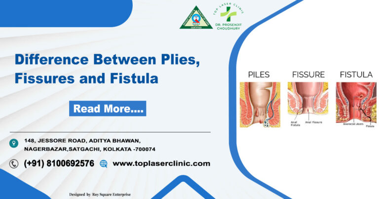
Fissures and Fistulas

The realm of gastrointestinal health often throws curveballs our way, and two common players in this complex arena are anal fissures and fistulas. These conditions, though seemingly similar, have distinct characteristics that necessitate individualized attention. In this blog post, we’ll delve into the nuances of anal fissures and fistulas, shedding light on their causes, symptoms, and treatment options.

Demystifying Anal Fissures:

Definition:

An anal fissure is akin to a small rift or tear in the delicate lining of the anal canal, causing discomfort and pain, particularly during bowel movements.

Culprits Behind the Fissure:

  • Constipation: Straining during bowel movements can lead to fissure formation.
  • Diarrhea: Frequent episodes of loose stools can irritate the anal tissue.
  • Trauma: Physical injury, including rough wiping or the insertion of foreign objects, may contribute to fissures.

Decoding Symptoms:

  1. Pain during bowel movements.
  2. Presence of bright red blood on toilet paper or in the toilet bowl.
  3. An itching or burning sensation in the anal region.

Remedies in the Arsenal:

  • Dietary adjustments: Increasing fiber intake to alleviate constipation.
  • Sitz baths: Soaking the anal area in warm water to facilitate healing.
  • Topical medications: Creams or ointments to reduce pain and inflammation.

Unraveling the Enigma of Anal Fistulas:

Definition:

An anal fistula, in contrast, manifests as a small tunnel connecting the anal canal to the skin near the anus. This unusual passageway typically stems from an infection in an anal gland.

The Architects of Fistulas:

  • Anal abscess: An untreated abscess can evolve into a fistula.
  • Crohn’s disease: Inflammatory bowel conditions can heighten the risk of fistula development.
  • Infections: Bacterial infections in anal glands can pave the way for fistula formation.

Clues in the Symptoms:

  1. Persistent or recurrent anal abscesses.
  2. Discharge of pus or blood from an opening near the anus.
  3. Pain and swelling in the anal area.

Strategies for Resolution:

  • Incision and drainage: Opening and draining abscesses to promote healing.
  • Fistulotomy: A surgical procedure to remove the fistula tract.
  • Seton placement: The introduction of a thin material to facilitate fistula drainage and healing.

In navigating the maze of anal fissures and fistulas, awareness is paramount. Recognizing the signs, understanding the causes, and seeking timely medical intervention can pave the way for effective management and a quicker road to recovery. Remember, your digestive health is a critical aspect of your overall well-being, and addressing these issues promptly ensures a smoother journey to a healthier you.

Differences between Anal Fissures and Fistulas:

Anal fissures and fistulas are two distinct conditions affecting the anal region, but they are often confused due to some similarities in symptoms. Here are the key differences between anal fissures and fistulas:

Anal Fissure: An anal fissure is a small tear or cut in the lining of the anal canal, typically near the opening of the anus.

Anal Fistula: An anal fistula is an abnormal tunnel-like tract that forms between the anal canal and the skin near the anus. It usually develops as a result of an infection or abscess in the anal glands.

Causes:

  • Anal Fissure: Fissures often result from trauma to the anal tissue, such as during bowel movements, especially in cases of constipation or diarrhea.
  • Anal Fistula: Fistulas commonly arise from an untreated anal abscess, which is a collection of pus that can form in the anal glands. Conditions like Crohn’s disease or infections can also contribute to fistula formation.

Location:

Anal Fissure: Typically located in the lining of the anal canal, near the surface of the anus.

Anal Fistula: Involves a tunnel that extends from the anal canal to the skin around the anus.

Symptoms:

Anal Fissure: Symptoms include pain during bowel movements, bright red blood on toilet paper or in the toilet bowl, and an itching or burning sensation in the anal area.

Anal Fistula: Symptoms include persistent or recurrent anal abscesses, discharge of pus or blood from an opening near the anus, and pain and swelling around the anal area.

Treatment:

Anal Fissure: Treatment may include dietary changes (increasing fiber), sitz baths (soaking the anal area in warm water), and topical medications (to reduce pain and inflammation).

Anal Fistula: Treatment may involve incision and drainage of abscesses, fistulotomy (surgical removal of the fistula tract), or seton placement (using a thread-like material to aid in draining and healing).

Chronicity:

Anal Fissure: Often acute and can resolve with conservative measures.

Anal Fistula: Tends to be a chronic condition and may require surgical intervention for resolution.

Understanding these differences is crucial for accurate diagnosis and appropriate treatment. If you suspect you have either of these conditions, it’s essential to consult with a healthcare professional for a proper evaluation and tailored treatment plan.

Can Fissures Transform into Fistulas?

Embarking on a journey through the intricacies of anal health, it’s essential to address a common question that often arises: Can anal fissures evolve into fistulas? Now, we’ll explore the relationship between these two conditions, understanding the factors that may contribute to this transition and the importance of early intervention.

Anal Fissures: A Brief Overview

Before delving into the potential transformation, let’s revisit what anal fissures entail. These are small tears or cuts in the lining of the anal canal, often caused by trauma during bowel movements, constipation, or diarrhea. Symptoms include pain, bleeding, and discomfort, typically treatable with conservative measures such as dietary changes and topical medications.

The Bridge Between Fissures and Fistulas:

Untreated Fissures:
  • Persistent Trauma: Anal fissures, if left untreated, may expose the surrounding tissue to persistent trauma during bowel movements.
  • Chronic Inflammation: The ongoing irritation and inflammation in the anal region can create an environment conducive to more severe complications.
Infection and Abscess Formation:
  • Bacterial Invasion: Prolonged trauma and inflammation can make the anal area susceptible to bacterial invasion.
  • Abscess Development: In some cases, bacterial infections can lead to the formation of anal abscesses, collections of pus in the anal glands.
Transitioning from Abscess to Fistula:

Natural Progression

  • Abscess Evolution: If anal abscesses remain untreated, they can evolve into more complex structures known as fistulas.
  • Tunnel Formation: The abscess may create a tunnel-like tract connecting the anal canal to the skin near the anus, resulting in the development of an anal fistula.
Underlying Conditions
  • Inflammatory Bowel Disease: Conditions like Crohn’s disease, which involve chronic inflammation in the digestive tract, can increase the likelihood of fistula development.
  • Immunocompromised State: Individuals with weakened immune systems may be more prone to complications, including the progression from fissures to fistulas.
The Importance of Early Intervention:

Recognizing the signs of anal fissures and seeking prompt medical attention is crucial to prevent potential complications. Early treatment can often resolve fissures, preventing them from evolving into more complex conditions like fistulas.

Seeking Professional Guidance:

If you’re experiencing persistent symptoms, including pain, bleeding, or swelling in the anal area, consulting a healthcare professional is essential. A timely diagnosis and appropriate treatment can help manage fissures effectively, reducing the risk of complications.

While anal fissures, if left unattended, can contribute to the development of anal fistulas, the key lies in early intervention. Understanding the progression and seeking professional guidance empower individuals to take control of their anal health, ensuring a smoother path to recovery and overall well-being. Remember, your health is a priority, and addressing concerns promptly is a crucial step in maintaining a balanced and comfortable life.

Sit Smart, Live Healthy: Embrace the Best Sitting Position for Varicose Vein Relief!

what-are-varicose-veins

Millions of people worldwide suffer from the widespread vascular ailment known as varicose veins. Despite their prevalence, many individuals may not fully understand what varicose veins are, their causes, symptoms, and available treatment options. In this blog post, we will delve into the world of varicose veins, providing valuable insights to increase awareness and promote a better understanding of this condition.

What are Varicose Veins?

Varicose veins are enlarged, swollen veins that usually appear blue or dark purple, often twisted and bulging. They most commonly occur in the legs and are the result of weakened or damaged valves within the veins. These valves, which normally prevent blood from flowing backward, may fail to function properly, causing blood to pool and veins to become enlarged.

Causes of Varicose Veins:

Heredity:Genetics: The chance of getting varicose veins is higher in families where the condition has already occurred.

Age: As individuals age, the veins may lose elasticity, leading to weakened valves.

Gender: Women are more likely to develop varicose veins, especially during pregnancy and hormonal changes.

Prolonged Standing or Sitting: Jobs or lifestyles that involve long periods of standing or sitting can contribute to the development of varicose veins.

Obesity: Excess weight puts additional pressure on the veins, potentially causing valve dysfunction.

Symptoms of Varicose Veins:

Visible, bulging veins: The most obvious sign of varicose veins is the appearance of swollen, twisted veins.

Aching or throbbing pain: Many individuals with varicose veins experience discomfort or pain in the affected areas.

Swelling and heaviness: Legs may feel heavy or swollen, particularly after extended periods of standing.

Skin changes: The skin around varicose veins may become discolored, dry, or itchy.

Treatment Options:

Lifestyle Changes: Regular exercise, maintaining a healthy weight, and avoiding prolonged periods of sitting or standing can help alleviate symptoms.

Compression Stockings: These special stockings apply pressure to the legs, promoting better blood flow and reducing swelling.

Sclerotherapy: A minimally invasive procedure involving the injection of a solution into the affected veins, causing them to collapse and fade.

Endovenous Laser Treatment (EVLT): A laser is used to close off the affected vein, redirecting blood flow to healthier veins.

Surgery: In some extreme cases, surgical procedures such as vein stripping or ligation are recommended.

Understand the best sitting position for varicose veins:

best-sitting-position-for-varicose-veins

For individuals dealing with varicose veins, choosing the right sitting position is crucial to alleviate symptoms and prevent further discomfort. In this blog post, we will explore the best sitting positions that can contribute to varicose vein relief and overall leg health.

Sit with Legs Elevated : Elevating your legs while sitting can help reduce the pressure on your veins and improve blood circulation. If possible, prop your legs up on a stool or another surface, ensuring that they are above the level of your heart. This position encourages blood to flow back towards the heart, reducing the likelihood of pooling in the veins.

Cross-Legged Position : Sitting cross-legged on the floor or with one ankle resting on the opposite knee can promote better circulation. This position helps in reducing pressure on the veins and prevents blood from pooling in the lower extremities.

Use a Footrest : Elevating your feet with a footrest or a small stool while sitting at a desk or table can improve blood flow and reduce strain on your veins. Ensure that your knees are bent at a 90-degree angle for optimal comfort.

Avoid Prolonged Sitting : Regardless of the position, it’s essential to avoid sitting for long periods continuously. Take breaks, stand up, stretch, and walk around periodically to keep the blood flowing and prevent stagnant circulation.

Sit on an Exercise Ball : Opting for an exercise ball as your chair promotes constant micro-movements, engaging the muscles in your legs and encouraging blood circulation. This dynamic sitting option can also help improve posture, reducing the strain on your veins.

Proper Ergonomics : If you spend significant time at a desk, ensure your workstation is set up with proper ergonomics. Adjust the height of your chair, desk, and computer to promote good posture and minimize stress on your legs.

Use Supportive Cushions : Consider using cushions or pillows to support your lower back and promote a more comfortable sitting position. This helps maintain good posture and reduces pressure on your veins.

While sitting is an inevitable part of our daily lives, adopting the right sitting positions can significantly contribute to varicose vein relief. Whether it’s elevating your legs, using supportive furniture, or incorporating regular breaks, these small adjustments can make a big difference in maintaining healthy blood circulation and preventing further discomfort associated with varicose veins. Always consult with a healthcare professional for personalized advice and additional strategies to manage varicose veins effectively.

Best Exercises for Varicose Vein Relief:

exercises-for-varicose-vein-relief

Now, we’ll explore some of the best exercises for varicose vein relief, focusing on activities that promote improved circulation, strengthen leg muscles, and contribute to overall vascular health.

Walking: Walking is a low-impact, accessible exercise that benefits overall cardiovascular health and helps pump blood from the legs back to the heart. Try to walk briskly for at least half an hour everyday. Consider incorporating intervals of faster walking to further enhance blood circulation.

Cycling:   Cycling is an excellent option for those seeking a low-impact exercise that engages the leg muscles. Whether on a stationary bike or outdoors, cycling promotes blood flow and strengthens the calf muscles, which play a key role in supporting healthy veins.

Swimming:Swimming and water aerobics are ideal for individuals with varicose veins as they provide resistance without putting stress on the joints. The buoyancy of water reduces the impact on the legs while working various muscle groups and promoting circulation.

Leg Elevation Exercises:Lie on your back and elevate your legs against a wall, allowing blood to flow back towards the heart. This simple exercise helps reduce swelling and pressure in the veins. Hold the position for 10-15 minutes, gradually increasing the duration as your comfort allows.

Calf Raises: Calf raises strengthen the muscles in the lower legs, improving blood circulation. With your feet hip-width apart, stand up, come up onto your toes, and then come back down. Repeat 15-20 times, gradually increasing the repetitions as your strength improves.

Ankle Flexes and Rotations: Sit or lie down and move your ankles in circular motions and flex them back and forth. This helps stimulate blood flow and prevents blood from pooling in the lower extremities.

Yoga: Certain yoga poses, such as legs-up-the-wall pose and inversions, can help promote blood circulation and alleviate symptoms associated with varicose veins. Consult with a yoga instructor to ensure proper form and technique.

Resistance Training:  Incorporating resistance training, especially focused on the lower body, can help build muscle strength. Exercises like squats and lunges engage multiple muscle groups, supporting overall leg health.

Regular exercise is a key component of managing varicose veins, promoting blood circulation, and strengthening the muscles that play a role in vascular health. By incorporating these exercises into your daily life, you can take proactive steps towards maintaining healthy legs and managing varicose veins effectively.

However, before starting an exercise routine for varicose veins, it’s crucial to consider several factors to ensure your safety and maximize the benefits. Here are some important things to keep in mind:

Consultation with Healthcare Professional:

Before beginning any exercise program, consult with your healthcare provider, especially if you have pre-existing medical conditions or concerns. They can assess your specific situation and provide personalized recommendations based on your health status.

Type of Varicose Veins:

Varicose veins vary in severity and location. The type and intensity of exercises may need to be tailored to your specific condition. Your healthcare provider can guide you on the most suitable exercises based on the location and severity of your varicose veins.

Avoid High-Impact Activities:

High-impact exercises, such as running or jumping, may exacerbate the symptoms of varicose veins. Choose low-impact activities like walking, cycling, or swimming to minimize stress on your veins and joints.

Compression Garments:

Consider wearing compression stockings or sleeves during exercise. These garments help improve blood circulation and reduce swelling in the legs. Your healthcare provider can recommend the appropriate level of compression for your condition.

Gradual Progression:

Exercise for a longer period of time and with more intensity after starting off gently. Activities that are sudden or severe can put strain on your veins and make you more uncomfortable. Pay attention to your body’s signals and move at a comfortable speed.

Proper Warm-Up and Cool Down:

Begin each exercise session with a proper warm-up to prepare your muscles and circulatory system. Cooling down afterward helps prevent muscle stiffness and promotes healthy blood circulation.

Hydration:

Stay well-hydrated before, during, and after exercise. Proper hydration supports overall vascular health and helps maintain optimal blood flow.

Avoid Prolonged Standing or Sitting:

Break up long periods of sitting or standing with short walks and stretches. Prolonged periods of immobility can contribute to poor circulation and worsen varicose vein symptoms.

Listen to Your Body:

Pay heed how your body responds to exercise. If you experience pain, swelling, or discomfort, modify your routine or consult with your healthcare provider for guidance.

Incorporate Leg Elevation:

After exercising, elevate your legs for a short period. This helps in reducing swelling and aids in the venous return of blood to the heart.

Choose Appropriate Footwear:

Wear comfortable, supportive shoes, especially if your exercise routine involves walking or other weight-bearing activities. Proper footwear can promote good posture and reduce strain on your legs.

Regular Check-ups:

Schedule regular follow-up appointments with your healthcare provider to monitor the progress of your varicose vein management. They can adjust your exercise plan as needed based on your health status.

Remember, the goal of exercising for varicose veins is to improve blood circulation, strengthen muscles, and reduce symptoms. By taking these considerations into account and working closely with your healthcare provider, you can create a safe and effective exercise plan tailored to your individual needs.

Varicose veins are a common vascular condition that can cause discomfort and affect the quality of life for those who experience them. Understanding the causes, symptoms, and treatment options, suitable exercises and proper sitting position is crucial for managing and preventing complications associated with varicose veins. If you suspect you have varicose veins or are experiencing related symptoms, consult with a healthcare professional for a thorough evaluation and personalized treatment plan.

Understanding do mammograms hurt ?

what-is-mammogram

Mammogram

In the realm of women’s health, the mammogram stands out as a crucial diagnostic tool, playing a pivotal role in early detection and prevention of breast cancer. This non-invasive screening procedure has been a cornerstone in the fight against breast cancer, saving lives and empowering women to take charge of their well-being. In this blog post, we’ll delve into the significance of mammograms, shedding light on their importance, procedure, and the conception that do mammograms hurt and the impact they have on women’s health.

The Importance of Mammograms:

Mammograms are specialized X-ray examinations designed to capture detailed images of the breast tissue. The primary purpose is to identify any abnormalities, such as lumps or masses, that may be indicative of breast cancer. Early detection through mammography significantly improves the chances of successful treatment, underlining the importance of regular screenings, particularly for women aged 40 and above.

Early Stage Breast Cancer Detection:

One of the key benefits of mammograms is their ability to detect breast cancer at an early stage, often before symptoms manifest. Early detection allows for prompt intervention, increasing the range of treatment options available and improving the likelihood of a positive outcome. By identifying abnormalities when they are still small and localized, mammograms contribute to more effective and less invasive treatment strategies.

The Mammogram Procedure:

Understanding the mammogram procedure can alleviate concerns and encourage women to prioritize regular screenings. During a mammogram, the breast is gently compressed between two plates of the mammography machine. While this compression may cause temporary discomfort, it is essential for obtaining clear and detailed images. The entire process is relatively quick, typically lasting around 20 minutes, and the discomfort is brief compared to the potential benefits of early cancer detection.

Types of Mammogram:

do-mammograms-hurt

Understanding the various types of mammograms available can empower individuals to make informed decisions about their breast health. In this blog post, we’ll explore the different types of mammograms and do mammograms hurt, shedding light on their purposes, procedures, and benefits.

Screening Mammograms: The Foundation of Prevention

  • Purpose: Designed for women without any breast cancer symptoms, screening mammograms aim to detect cancer in its early stages when treatment is most effective.
  • Procedure: Patients stand in front of an X-ray machine, and each breast is compressed for a few seconds to capture detailed images.
  • Benefits: Early detection and increased chances of successful treatment.

Diagnostic Mammograms: Investigating Suspicious Findings

  • Purpose: Conducted when a patient exhibits symptoms like a lump, pain, or discharge, diagnostic mammograms provide a closer look to identify the cause.
  • Procedure: Similar to screening mammograms but with additional views for a more in-depth analysis.
  • Benefits: Pinpointing the nature of abnormalities for accurate diagnosis.

3D Mammography (Tomosynthesis): Revolutionizing Detection

  • Purpose: Offers a three-dimensional view of the breast, reducing the likelihood of false positives and improving cancer detection rates.
  • Procedure: Similar to traditional mammograms but captures multiple images at different angles.
  • Benefits: Enhanced clarity, especially in dense breast tissue, and reduced callback rates.

Digital Mammography: Advancing Image Quality

  • Purpose: Utilizes digital technology for image capture and storage, providing clearer images and facilitating easier sharing with healthcare providers.
  • Procedure: Similar to traditional mammograms but with digital receptors instead of X-ray film.
  • Benefits: Improved image quality, quicker results, and the ability to manipulate images for better analysis.

Contrast-Enhanced Mammography (CEM): Spotlighting Abnormalities

  • Purpose: Involves the injection of a contrast agent to highlight blood flow in the breast, aiding in the detection of lesions that may not be visible on standard mammograms.
  • Procedure: Combined with a standard mammogram, the contrast agent enhances the visibility of abnormalities.
  • Benefits: Improved detection of abnormalities, especially in high-risk patients.

Frequency of Mammograms:

The frequency of mammograms depends on various factors, including age, family history, and individual risk factors. Generally, women are encouraged to begin regular mammograms at the age of 40 and continue to do so annually or biennially. For those with a higher risk of breast cancer, healthcare providers may recommend more frequent screenings or additional imaging techniques.

Bilateral Mammogram:

Bilateral mammograms involve the imaging of both breasts, capturing detailed X-ray images to identify any abnormalities or potential signs of breast cancer. Unlike unilateral mammograms, which focus on a single breast, bilateral mammograms provide a more complete picture of the breast tissue. This comprehensive approach enhances the accuracy of detecting abnormalities, ensuring a more thorough assessment of breast health.

The Procedure:

The process of undergoing a bilateral mammogram is similar to a unilateral mammogram. The patient stands in front of the mammography machine, and each breast is compressed between two plates to obtain high-quality images. While the compression might be momentarily uncomfortable, it is necessary to ensure optimal image clarity. The entire procedure typically takes around 20 to 30 minutes and is a relatively routine and non-invasive examination.

Advantages of Bilateral Mammograms:

advantages-of-bilateral-mammograms
  1. Increased Sensitivity: Bilateral mammograms have a higher sensitivity for detecting breast abnormalities compared to unilateral mammograms. This is crucial for early detection and prompt intervention.
  2. Comparative Analysis: With images from both breasts available, radiologists can make more accurate comparisons, distinguishing between normal variations and potential abnormalities.
  3. Risk Assessment: Bilateral mammograms aid in assessing the overall risk of breast cancer, especially for individuals with a family history or other risk factors.This information can guide personalized screening and prevention strategiesStrategies for preventive and customised screening can be informed by this data..
  4. Comprehensive Evaluation: As breast tissue varies, having a bilateral view ensures a more comprehensive evaluation of the entire breast anatomy, including the axillary region.

Bilateral mammograms are a vital tool in the early detection of breast cancer, offering a more thorough examination and increasing the chances of identifying abnormalities at their earliest, most treatable stages. Regular screenings, along with maintaining open communication with healthcare providers, empower individuals in taking charge of their breast health. Remember, early detection saves lives, and bilateral mammograms are a significant step forward in this ongoing battle against breast cancer.

Empowering Women Through Knowledge:

Knowledge is a powerful tool in the realm of health, and understanding the significance of mammograms empowers women to take control of their breast health. By prioritizing regular screenings and embracing early detection, women can actively contribute to their overall well-being and play a proactive role in the prevention and management of breast cancer.

Do mammograms hurt:

Despite their life-saving potential, many women are hesitant to undergo this screening due to concerns about discomfort or pain. In this blog post that do mammograms hurt, we aim to debunk the myth surrounding mammograms and explore the reality of the experience that do mammograms hurt.

Body:

  1. Understanding the Importance of Mammograms: Begin by emphasizing the importance of mammograms in detecting breast cancer at its earliest, most treatable stage. Highlight statistics and success stories to reinforce the life-saving impact of regular screenings.
  2. Setting Realistic Expectations: Acknowledge that every woman’s experience may vary, but it’s crucial to set realistic expectations. Most women report minor discomfort rather than significant pain during the procedure. Educate readers on the purpose of compression during mammograms and how it aids in obtaining clearer images.
  3. The Mammogram Process: Break down the mammogram process step by step, from check-in to the actual imaging. Explain how the compression of the breast is brief and necessary for accurate results. Emphasize that the discomfort lasts only for a few seconds during each compression.
  4. Tips for a Comfortable Experience: Provide practical tips for minimizing discomfort during a mammogram. This may include scheduling the appointment at a specific time of the menstrual cycle when breasts are less tender, using relaxation techniques, or taking over-the-counter pain relievers before the procedure.
  5. Patient Testimonials: Share positive experiences from women who have undergone mammograms. Include stories of early cancer detection and successful treatments that resulted from routine screenings. Real-life testimonials can help alleviate fears and inspire confidence in the screening process.
  6. Addressing Common Concerns: Acknowledge specific concerns that women may have, such as fear of radiation exposure or anxiety about receiving abnormal results. Provide factual information to dispel myths and ease these worries.
  7. Alternative Screening Options: Discuss alternative breast cancer screening methods for women who may have a particularly low pain tolerance or other medical considerations. Highlight the importance of consulting with healthcare professionals to determine the most suitable screening approach.

In conclusion, emphasize that the potential discomfort associated with mammograms should not deter women from undergoing this critical screening. The benefits of early detection far outweigh the momentary discomfort, and with proper knowledge and preparation, women can approach mammograms with confidence and a commitment to their long-term health.

In the journey towards optimal health, mammograms stand as a beacon of hope and empowerment. As a widely accessible and effective tool, they enable early detection, fostering positive outcomes in the battle against breast cancer. By embracing regular mammograms, women not only prioritize their health but also contribute to a collective effort to reduce the impact of breast cancer on individuals and communities alike. Remember, early detection saves lives, making mammograms an invaluable ally in the pursuit of a healthier and happier future.

Remember to encourage readers to consult with their healthcare providers if they have specific concerns or questions about mammograms tailored to their individual health needs.

The 3 Finger Test for Hernia: A Simple Guide to Self-Assessment

3-Finger-Test-for-Hernia

Many people may experience discomfort and worry due to hernias. The good news is that there’s a simple and quick self-assessment method known as the “3 Finger Test” that can help you identify potential signs of a hernia. In this blog post, we’ll explore what a hernia is, how the 3 Finger Test works, and what steps you can take if you suspect you may have a hernia.

Understanding Hernias:

Before delving into the 3 Finger Test, let’s briefly understand what a hernia is. An organ or tissue that pushes through a weak area in the surrounding muscle or connective tissue results in a hernia. Hiatal (upper stomach), umbilical (belly button), and inguinal (groyne) hernias are common varieties. Symptoms may include a visible bulge, pain, and discomfort, particularly during activities like lifting or coughing.

The 3 Finger Test:

The Three Finger Test is a physical examination method used by healthcare practitioners to identify the presence of hernias in patients. The test primarily focuses on the abdominal wall, aiming to detect any weakness or protrusion indicative of a hernia.
The 3 Finger Test is a straightforward self-examination that can be performed at home. Here’s how to do it:

Find a Comfortable Position

Start by lying down in a relaxed position. You can choose to lie on your back or your side, whichever is more comfortable for you.

Locate the Area of Concern:

Place three fingers of one hand on the area where you suspect a hernia might be present. This is typically in the abdominal or groin region.

Apply Gentle Pressure:

With the three fingers in place, apply gentle pressure to the area. Pay attention to any sensations such as pain, tenderness, or the feeling of a lump or bulge.

Cough or Strain:

While maintaining the pressure, cough or strain as if you were having a bowel movement. This action may cause a hernia to become more noticeable.

Purpose of the Three Finger Test:

  • Hernia Identification: The primary objective of the Three Finger Test is to identify the presence of hernias, particularly in the abdominal region. Hernias occur when an organ or tissue pushes through a weakened area of the surrounding muscle or connective tissue.
  • Non-Invasive Screening: Unlike some diagnostic procedures, the Three Finger Test is non-invasive, making it a valuable tool for initial screening before more advanced imaging or diagnostic tests are considered.

Interpreting the Results:

If you experience pain, tenderness, or detect a bulge during the 3 Finger Test, it’s essential to consult with a healthcare professional promptly. They can conduct a thorough examination, potentially including imaging tests, to confirm the presence of a hernia and determine the appropriate course of action.

Significance in Medical Diagnostics:

significance-in medical-diagnostic
  • Early Detection: Early detection of hernias is crucial for effective treatment. The Three Finger Test allows healthcare professionals to identify potential issues before they escalate, enabling timely intervention.
  • Cost-Effective: As a simple physical examination, the Three Finger Test is a cost-effective method for hernia screening compared to more complex imaging procedures.
  • Patient Comfort: Given its non-invasive nature, the test is generally well-tolerated by patients, contributing to a positive patient experience during the diagnostic process.

Seeking Professional Guidance:

While the 3 Finger Test can be a useful initial self-assessment, it’s crucial to remember that it is not a substitute for professional medical advice. If you suspect you have a hernia or experience any concerning symptoms, consult with a healthcare provider for a comprehensive evaluation.

The 3 Finger Test offers a simple and accessible way for individuals to perform an initial self-assessment for hernias. However, it’s important to emphasize the importance of seeking professional medical advice for a definitive diagnosis and appropriate treatment. Early detection and intervention can significantly contribute to effective hernia management and improved overall well-being.

Can a hernia surgery fail?

If the hernia surgery is done correctly, it does not come back and in some cases it may come back, then it is called a recurrent hernia.

How do you test for inguinal hernia with three fingers?

the fingers placed over the femoral region, the external inguinal ring, and the internal ring, have the patient cough, This is swelling which is locatedin any one of these areas may indicate a hernia.

How do you test for inguinal hernia with three fingers?

When we put fingers between the femoral region, the external inguinal ring, and the internal ring, if any swelling observed in any one of these areas may indicate a hernia.

Can you feel a hernia with your fingers?

By hiatal hernia test we can feel hernia with our fingers, by simply putting our fingers over the upper abdomen area and meanwhile deep inhaling can help us to find lump beneath our skin.

Is hernia a major problem?

Hernia is a very common disease but not a complicated one and can be cured with proper advice from a doctor.Since hernia does not go away on its own, it needs to be treatment properly.

Does food affect hernia?

If you have a hernia, you should avoid certain foods like oily or spicy food, Otherwise it takes a long time to reduce this disease.