Home Blog

Empowering Your Legs: The Role of Physiotherapy in Varicose Vein Management

Physiotherapy for Varicose Veins

Varicose veins are a common but often misunderstood vascular condition that affects a significant number of people worldwide. These swollen, twisted veins can cause discomfort and aesthetic concerns, impacting the quality of life for those affected. In this blog post, we will explore the role of physiotherapy in promoting vascular health, strengthening leg muscles, and enhancing overall well-being. However, prior to that we will delve into the causes, symptoms, and various treatment options available for managing varicose veins in short.

What are Varicose Veins?

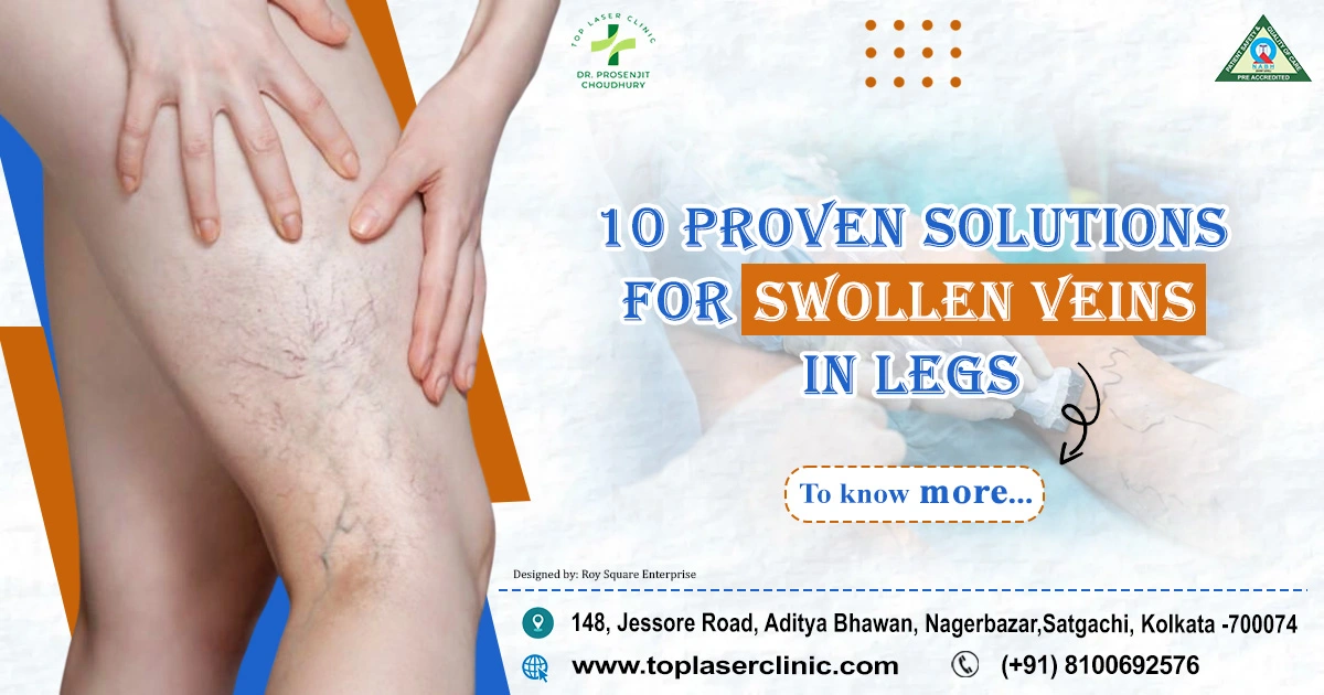
Larger, twisted veins that typically show up on the legs and feet are called varicose veins. They develop when the valves in the veins fail to function properly, causing blood to pool and exert increased pressure on the vein walls. This can result in the characteristic bulging and twisting of the veins that many people associate with varicose veins.

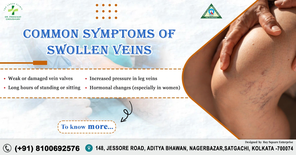
Causes of Varicose Veins:

Varicose veins can develop due to a number of circumstances, such as:

  • Genetics: Varicose veins are more common in families where the problem runs in the family.
  • Age: As people age, the elasticity of vein walls decreases, making them more prone to dilation and the development of varicose veins.
  • Gender: Women are more prone to develop varicose veins than men, partly due to hormonal changes during pregnancy and menopause.
  • Prolonged Standing or Sitting: Jobs that involve long periods of standing or sitting can impede blood flow, increasing the risk of varicose veins.
  • Obesity: Excess weight puts additional pressure on the veins, contributing to the development of varicose veins.

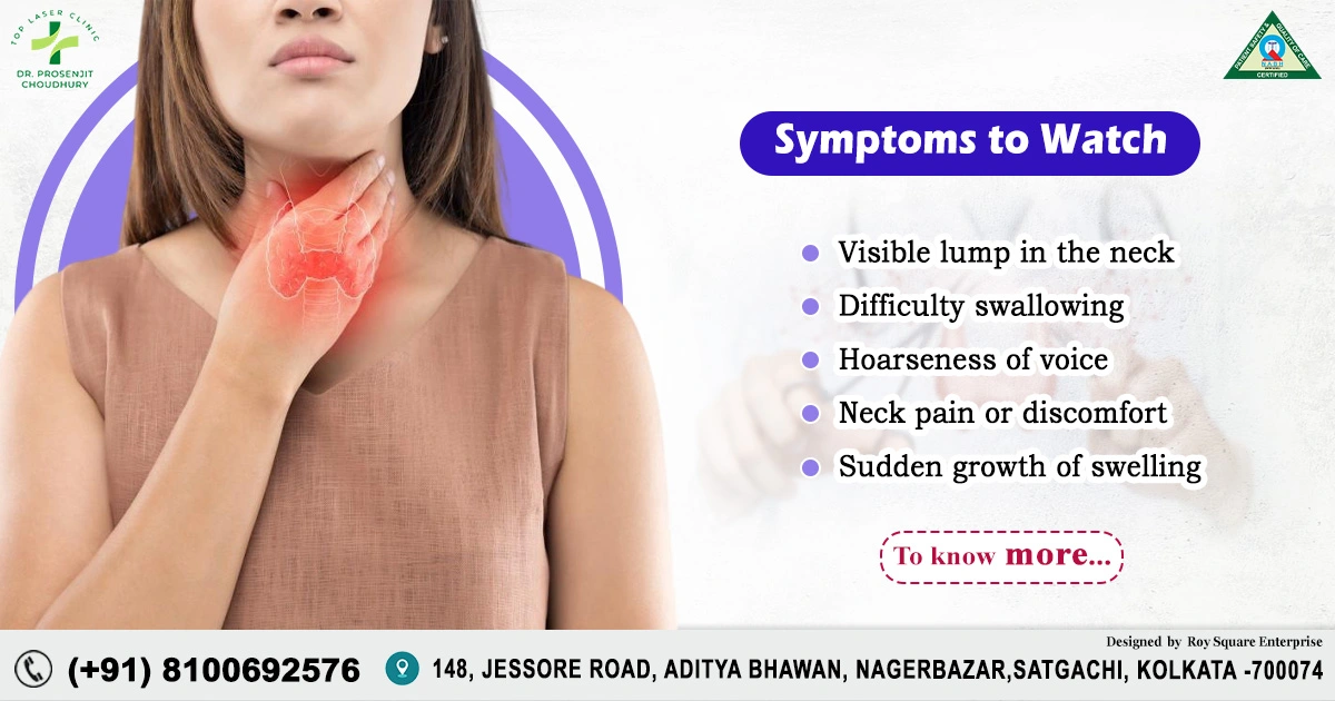
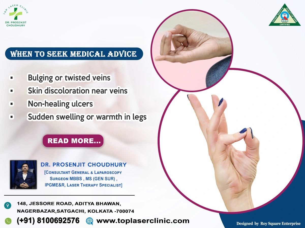
Symptoms of Varicose Veins:

Varicose veins may be asymptomatic for some individuals, while others may experience various symptoms, including:

  • Pain or Aching: Discomfort or aching in the affected area, especially after prolonged periods of standing or sitting.
  • Swelling: Swelling in the legs and ankles due to fluid build-up.
  • Itching and Burning: Skin irritation, itching, or a burning sensation around the affected veins.
  • Skin Changes: Changes in skin colour or the development of sores near the affected veins.
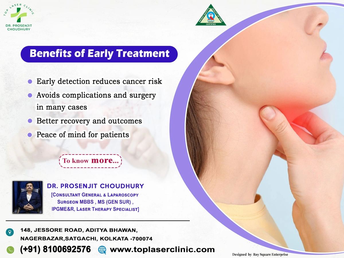
Treatment Options:

Several treatment options are available for managing varicose veins, ranging from lifestyle changes to medical interventions:

  • Lifestyle Modifications:Regular exercise to improve circulation.Weight management to reduce pressure on veins.Try to avoid prolonged periods of standing or sitting.
  • Compression Stockings:Wearing compression stockings to promote blood flow and reduce swelling.
  • Sclerotherapy:Injection of a solution into the affected veins to cause them to collapse and fade.
  • Endovenous Laser Treatment (EVLT):A minimally invasive procedure using laser energy to seal the affected veins.
  • Surgical Options:Vein stripping or ligation in severe cases.

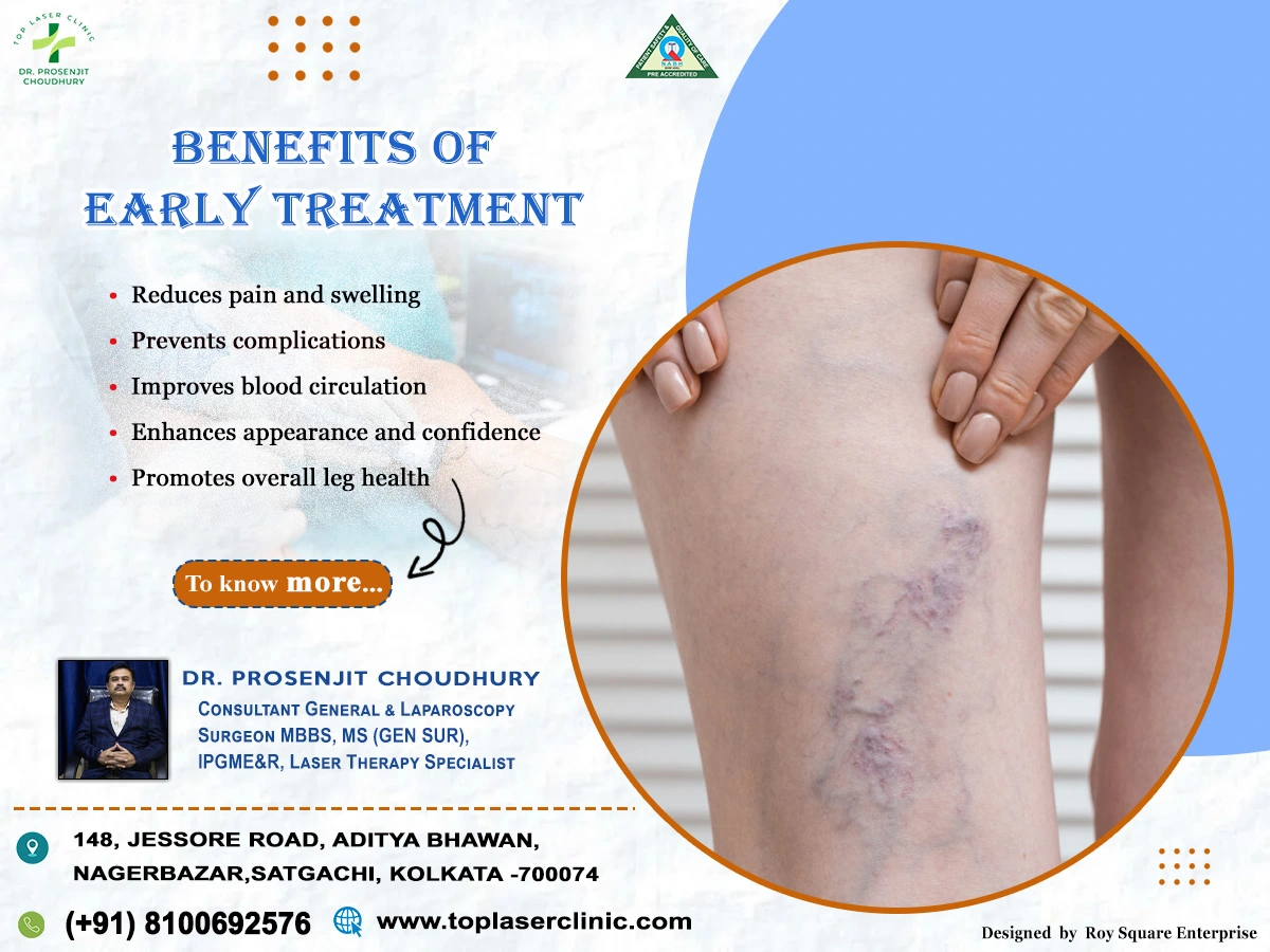
While various treatment options are available, physiotherapy stands out as a valuable and non-invasive approach to managing and alleviating the symptoms associated with varicose veins.

Understanding Physiotherapy for varicose veins

Physiotherapy, also known as physical therapy, involves the use of physical methods, exercises, and manual techniques to restore, maintain, and improve physical function. In the context of varicose veins, physiotherapy focuses on enhancing blood circulation, reducing swelling, and strengthening the muscles that support the venous system.

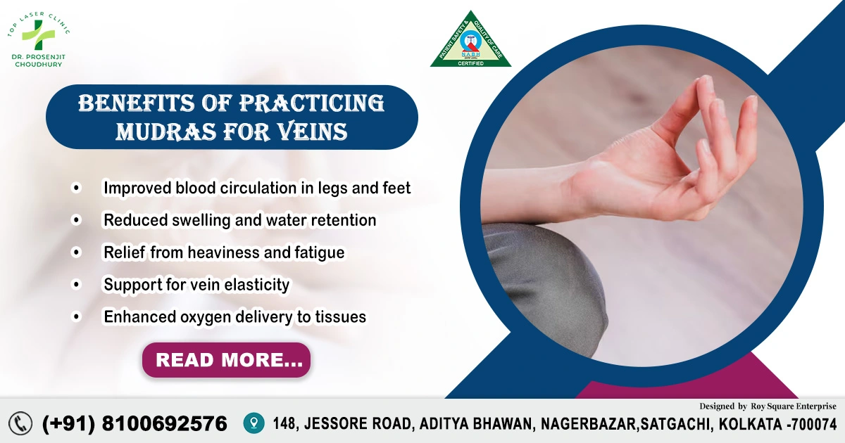
Benefits of Physiotherapy for Varicose Veins:

  • Improved Circulation:

Physiotherapy includes exercises and techniques that promote blood circulation, helping to reduce the pooling of blood in the affected veins. Improved circulation contributes to overall vascular health and minimizes the risk of complications.

  • Muscle Strengthening:

Physiotherapists design exercise programs to target specific muscle groups, particularly those in the legs. Strengthening these muscles helps support the venous system, reducing the pressure on veins and aiding in blood flow back to the heart.

  • Compression Therapy:

Physiotherapists may recommend compression stockings or bandages to help reduce swelling and improve blood circulation in the legs. Properly fitted compression garments can be a valuable component of a comprehensive physiotherapy plan.

  • Range of Motion Exercises:

Physiotherapy includes movements that promote flexibility and joint mobility. These exercises can be beneficial for individuals with varicose veins, preventing stiffness and discomfort associated with prolonged periods of inactivity.

  • Education and Lifestyle Modification:

Physiotherapists educate patients about lifestyle changes that can positively impact their vascular health. This may include advice on maintaining a healthy weight, staying active, and adopting ergonomic practices to reduce the risk of exacerbating varicose veins.

  • Manual Therapy Techniques:

Manual therapies such as massage and manual lymphatic drainage may be employed by physiotherapists to reduce swelling, alleviate pain, and enhance the overall well-being of individuals with varicose veins.

  • Posture Correction:

Correcting posture is crucial in managing varicose veins. Physiotherapists can assess and provide guidance on maintaining proper posture during various activities, reducing unnecessary strain on the venous system.

  • Collaboration with Healthcare Professionals:

Physiotherapy for varicose veins is often part of a collaborative approach involving various healthcare professionals, including vascular specialists. Physiotherapists work closely with patients to tailor treatment plans based on individual needs and the severity of the condition.

Physiotherapy emerges as a valuable and holistic approach in the management of varicose veins. By focusing on improving circulation, strengthening leg muscles, and promoting overall well-being, physiotherapy empowers individuals to take an active role in their vascular health. If you are dealing with varicose veins, consider consulting with a physiotherapist to develop a personalized plan that addresses your unique needs and enhances your quality of life.

Benefits of Massage for Varicose Veins:

  1. Improved Blood Circulation:

Massage helps stimulate blood flow, particularly in the affected areas. By applying gentle pressure and specific techniques, massage encourages blood to move through the veins more effectively, reducing the likelihood of blood pooling and congestion.

  • Reduced Swelling and Edema:

The gentle manipulation of tissues during a massage helps in draining excess fluid from the legs, reducing swelling (edema). This can provide significant relief to individuals experiencing discomfort and heaviness associated with varicose veins.

  • Relief from Pain and Discomfort:

Massage therapy can alleviate pain and discomfort associated with varicose veins by relaxing the muscles, reducing tension, and promoting a sense of well-being. Targeted massage techniques can specifically address areas with increased sensitivity.

  • Stimulation of Lymphatic Drainage:

Massage supports the lymphatic system, which plays a crucial role in removing waste and excess fluids from the body. By enhancing lymphatic drainage, massage contributes to the reduction of swelling and promotes a healthier vascular system.

  • Improved Muscle Function:

Varicose veins can lead to weakened muscles in the legs. Massage helps in toning and relaxing these muscles, improving their function and providing additional support to the venous system.

  • Enhanced Mental Well-being:

Beyond the physical benefits, massage has a positive impact on mental well-being. The relaxation induced by massage can help reduce stress and anxiety, which are factors that can contribute to the exacerbation of varicose veins symptoms.

Considerations Before Getting a Massage-

i. Consultation with a Healthcare Professional:Before undergoing massage therapy for varicose veins, it is advisable to consult with a healthcare professional, especially if you have any underlying health conditions.

ii. Choose a Qualified Massage Therapist:Opt for a licensed and experienced massage therapist who is knowledgeable about varicose veins. They can tailor the massage techniques to address your specific needs and ensure safety.

iii. Gentle Techniques:Inform your massage therapist about your varicose veins, and they can use gentle techniques to avoid putting excessive pressure on the affected areas.

Massage therapy offers a natural and soothing approach to managing the symptoms of varicose veins in the legs. By promoting better circulation, reducing swelling, and providing overall relief, massage can be a valuable addition to a comprehensive treatment plan. Always seek advice from your healthcare provider before starting any new therapeutic regimen, and enjoy the soothing benefits that massage can bring to your journey towards healthier, more comfortable legs.

Basic Information about Varicose Vein

Basic Information about Varicose Vein

Basic Information about Varicose Vein : Varicose veins are a common condition that affects millions of people worldwide. While they can occur in anyone, they are more common among women and typically affect the legs. Varicose veins occur when the valves in the veins weaken or malfunction, causing blood to pool and flow backward. This can lead to the veins becoming large, twisted and bulging. This condition can cause discomfort and pain, swelling, aching or throbbing in the affected area, and may even lead to more serious complications such as blood clots or ulcers.

Basic Information about Varicose Vein

If you are suffering from varicose veins, it is important to seek medical attention and consider treatment options. Ignoring the condition can lead to more severe symptoms, affect your quality of life and increase the chances of complications in the long run. There are various treatments available for varicose veins, including lifestyle changes, compression stockings, and medical procedures such as vein stripping or laser treatment. It is recommended that you consult with a qualified healthcare professional to determine the best course of action for your specific case.

Remember, varicose veins are a common and treatable condition that should not be ignored. If left untreated, it can lead to more serious complications that could affect your overall health and quality of life. Seek treatment from a qualified healthcare professional as soon as possible. Remember to maintain a healthy weight, exercise regularly, and avoid sitting or standing for prolonged periods to reduce your chances of developing varicose veins.

In summary, varicose veins are a common and treatable condition that can cause discomfort and serious complications if left untreated. It is important to seek medical attention and consider treatment options. Lifestyle changes such as maintaining a healthy weight, exercising regularly and avoiding sitting or standing for prolonged periods can help reduce the chances of developing varicose veins. Remember that seeking treatment for varicose veins is not only important for managing the condition but also can relieve discomfort and ensure a good quality of life. By adopting healthy habits and seeking treatment for varicose veins, you can improve your overall wellness and avoid the discomfort and complications associated with this condition. In conclusion, taking care of your cardiovascular health should be a priority in order to prevent varicose veins as well as other conditions like deep vein thrombosis. Maintaining healthy habits and seeking medical attention when necessary can greatly improve your overall wellbeing. Take action now to manage your varicose veins and improve your overall health. Don’t let varicose veins affect your quality of life. Incorporate healthy habits into your routine and consider treatment options if necessary to manage varicose veins and improve your overall wellness. Remember, prevention is always the best medicine. Always consult with your healthcare provider before making lifestyle changes or undergoing any medical treatments.

Thank you for taking the time to learn about varicose veins and how to manage them. Remember to prioritize your cardiovascular health and seek medical advice if you suspect the presence of varicose veins. Take care of your body and listen to its signals. Your body is a temple and it’s important to keep it healthy. Maintaining a healthy lifestyle is crucial for preventing and managing varicose veins, as well as other cardiovascular conditions. Making healthy choices and seeking regular medical check-ups can go a long way in ensuring your overall wellbeing. Stay active, eat a balanced diet and manage your stress levels to maintain optimal cardiovascular health.

Diet for Varicose Vein:

While there is no specific diet that can cure varicose veins, maintaining a healthy diet can help support overall vein health and manage the symptoms associated with varicose veins. Here are some dietary recommendations that may be beneficial:

  • 1. Fiber-Rich Foods: Consuming a diet high in fiber can help prevent constipation, which can contribute to increased pressure on the veins. Include foods such as whole grains, fruits, vegetables, legumes, and nuts in your diet.
  • 2. Antioxidant-Rich Foods: Antioxidants help protect the veins and improve overall circulation. Include foods rich in antioxidants, such as berries, citrus fruits, leafy greens, bell peppers, and dark chocolate.
  • 3. Omega-3 Fatty Acids: Omega-3 fatty acids have anti-inflammatory properties and can support cardiovascular health. Include sources of omega-3 fatty acids, such as fatty fish (salmon, mackerel, sardines), flaxseeds, chia seeds, and walnuts in your diet.
  • 4. Vitamin C: Vitamin C plays a role in collagen synthesis, which helps maintain the strength and elasticity of blood vessels. Include foods like citrus fruits, strawberries, kiwi, bell peppers, and broccoli to ensure an adequate intake of vitamin C.
  • 5. Potassium-Rich Foods: Potassium helps regulate fluid balance and can reduce water retention and swelling. Include foods like bananas, avocados, spinach, sweet potatoes, and tomatoes, which are good sources of potassium.
  • 6. Hydration: Stay adequately hydrated by drinking plenty of water throughout the day. Proper hydration helps maintain healthy blood circulation and can reduce the risk of blood clot formation. 7. Limit Salt Intake: High sodium intake can contribute to water retention and swelling. Reduce your consumption of processed and packaged foods, as they are often high in sodium. opt for fresh, whole foods and use herbs and spices to flavour your meals instead of salt.
  • 8. Maintain a Healthy Weight: Excess weight can put additional strain on your veins. Maintain a healthy weight through a balanced diet and regular exercise to reduce the pressure on your veins.

It’s important to note that while these dietary recommendations can support overall vein health and symptom management, they do not replace medical treatment or address the underlying causes of varicose veins.

Top 10 Incredible Benefits of Laparoscopic Surgery for Faster Healing | Dr. Prosenjit Choudhury

When it comes to surgery, every patient hopes for less pain, minimal scars, and faster recovery. With modern medical advancements, laparoscopic surgery — also known as keyhole surgery — has made all these possible.

According to Dr. Prosenjit Choudhury, Consultant General & Laparoscopy Surgeon, MBBS, MS (Gen Sur.), IPGME&R, and one of the best in laser & minimally invasive surgery at Top Laser clinic, Kolkata, laparoscopic surgery is transforming the way surgeries are performed today. It offers high precision, faster healing, and minimal post-surgical discomfort.

In this detailed blog, let’s explore what laparoscopic surgery is, how it works, its benefits, and why Top Laser Clinic, Kolkata is one of the best centres for these advanced procedures.

1. What is Laparoscopic Surgery?

Laparoscopic surgery is a minimally invasive surgical technique that allows doctors to perform operations using tiny incisions instead of large cuts.

A small camera (laparoscope) and specialized instruments are inserted through these small incisions. The camera projects real-time images onto a screen, allowing the surgeon to operate with great accuracy.

Dr. Prosenjit Choudhury, Consultant General & Laparoscopy Surgeon at Top Laser Clinic, Kolkata, explains that this method ensures minimal blood loss, less pain, and quicker recovery — making it the preferred choice for most abdominal and general surgeries today.

2. Symptoms Indicating You May Need Laparoscopic Surgery

Laparoscopic surgery is used for a wide range of conditions, especially those affecting the abdomen or pelvis. You might need evaluation for laparoscopic treatment if you experience:

  • Persistent abdominal pain
  • Swelling or lump in the abdomen
  • Gallstones or gallbladder pain
  • Appendix pain (appendicitis)
  • Hernia bulge or discomfort
  • Unexplained digestive issues
  • Recurrent infections in the abdomen or pelvis

If you notice any of these symptoms, it’s best to consult Dr. Prosenjit Choudhury, best general surgeon in Kolkata, for a detailed diagnosis and treatment plan.

3. Types of Laparoscopic Surgeries

Laparoscopic surgery is not limited to one condition — it’s used for multiple medical issues.

Here are some common types of laparoscopic procedures performed at Top Laser Clinic, Kolkata:

  1. Laparoscopic Appendectomy – Removal of the appendix.
  2. Laparoscopic Cholecystectomy – Removal of the gallbladder (for gallstones).
  3. Laparoscopic Hernia Repair – Fixing hernias using mesh support.
  4. Laparoscopic Diagnostic Surgery – For identifying unknown abdominal issues.
  5. Laparoscopic Hysterectomy – Removal of the uterus (for female patients).
  6. Laparoscopic Bariatric Surgery – Weight-loss surgery for obesity.
  7. Laparoscopic Adhesiolysis – Removing internal scar tissues.

These procedures are known for faster healing and minimal post-operative pain.

4. Causes for Needing Laparoscopic Surgery

There are several medical causes that may lead to the need for laparoscopic intervention, such as:

  • Chronic or acute abdominal infections
  • Gallbladder stones or inflammation
  • Appendix infection
  • Hernia formation
  • Uterine fibroids or cysts
  • Obesity-related complications
  • Tumours or abnormal growths in the abdomen

Early diagnosis and timely treatment using laparoscopic methods prevent complications and promote faster recovery.

5. Risk Factors for Abdominal or Pelvic Disorders

Certain factors can increase the risk of developing abdominal or pelvic diseases that may need laparoscopic surgery, including:

  • Poor diet and obesity
  • Family history of gallbladder or hernia problems
  • Lack of exercise
  • Smoking or alcohol use
  • Previous abdominal surgery
  • Age-related muscle weakness

Knowing your risk factors can help you take preventive steps and consult a specialist early.

6. Diagnosis before Laparoscopic Surgery

Before recommending surgery, a detailed diagnosis is done to ensure accuracy and safety. At Top Laser Clinic, Kolkata, diagnosis includes:

  • Physical Examination – Checking for tenderness, swelling, or hernia.
  • Ultrasound / CT scan – To get a clear image of internal organs.
  • Blood Tests – To detect infection or inflammation.
  • Endoscopic Examination – In some cases, to look inside the abdomen.

Dr. Prosenjit Choudhury, Consultant General & Laparoscopy Surgeon, ensures every patient undergoes a thorough evaluation before surgery for the best outcome.

7. Treatment Options

1. Laparoscopic (Minimally Invasive) Surgery

  • Involves small incisions and a camera for precision.
  • Faster healing, minimal scarring, and less pain.
  • Shorter hospital stay and quicker return to normal life.

2. Open Surgery

  • Requires a larger incision.
  • Usually recommended for complex or emergency cases.

3. Non-Surgical Management

  • For very mild or early cases, medication or therapy may help.

At Top Laser Clinic, patients are guided carefully on the best approach depending on their specific condition.

8. Recovery after Laparoscopic Surgery

One of the biggest advantages of laparoscopic surgery is faster recovery compared to traditional open surgery.

Typical Recovery Timeline:

  • Discharge: Within 1–2 days after surgery
  • Return to work: Within 5–7 days
  • Full recovery: Within 2–4 weeks

Tips for Smooth Recovery:

  • Follow your surgeon’s dietary advice.
  • Avoid lifting heavy objects for 2–3 weeks.
  • Keep the incision site clean.
  • Attend follow-up check-ups regularly.

Patients treated by Dr. Prosenjit Choudhury often experience minimal post-surgery pain and resume normal life quickly.

9. Possible Complications (Though Rare)

Laparoscopic surgery is extremely safe in expert hands, but minor complications can rarely occur, such as:

  • Temporary pain at the incision site
  • Mild swelling or bruising
  • Infection (very rare)
  • Bleeding or reaction to anaesthesia

At Top Laser Clinic, Kolkata, strict hygiene and advanced technology ensure maximum patient safety.

Top 10 Benefits of Laparoscopic Surgery

  1. Smaller incisions and minimal scarring
  2. Less postoperative pain
  3. Reduced risk of infection
  4. Shorter hospital stay
  5. Quicker recovery and return to work
  6. Minimal blood loss
  7. Better cosmetic results
  8. Enhanced surgical precision
  9. Lower healthcare costs in the long run
  10. Higher patient satisfaction

These benefits make laparoscopic surgery a top choice among patients — and Dr. Prosenjit Choudhury, best general surgeon in Kolkata, has mastered this modern approach for outstanding outcomes.

10. Top 10 Frequently Asked Questions (FAQs)

1. What is laparoscopic surgery?
It’s a modern, minimally invasive surgery that uses small cuts and a camera for high precision.

2. Is laparoscopic surgery safe?
Yes, it’s one of the safest surgical options available today.

3. How long does it take to recover?
Most patients recover fully within 2–4 weeks.

4. Does it cause much pain?
No, pain is significantly less compared to open surgery.

5. Will I have visible scars?
Only small marks remain, which fade over time.

6. Is hospitalization needed?
Usually just 1–2 days for most laparoscopic procedures.

7. Can elderly patients undergo laparoscopic surgery?
Yes, if medically fit, elderly patients recover well with this technique.

8. What are the risks involved?
Minor risks include temporary pain or mild infection, which are easily manageable.

9. How do I prepare for the surgery?
Follow your surgeon’s fasting and medication guidelines before surgery.

10. Where can I get the best laparoscopic surgery in Kolkata?
At Top Laser Clinic, Kolkata, under the expert care of Dr. Prosenjit Choudhury, Consultant General & Laparoscopy Surgeon, MBBS, MS (Gen Surg), IPGME&R, best in laser & minimally invasive surgery.

Why Choose Top Laser Clinic, Kolkata?

Top Laser Clinic is one of Kolkata’s leading centres for advanced laser and laparoscopic surgeries, providing high-quality, patient-focused care.

Here’s Why Patients Trust Us:

  • Expertise of Dr. Prosenjit Choudhury, best general surgeon in Kolkata, renowned for his precision and experience in laser and minimally invasive surgery.
  • Modern operation theatres with cutting-edge laparoscopic technology.
  • Fast, painless, and scar-minimizing treatments.
  • Personalized pre and post-surgery care.
  • Affordable, transparent pricing with excellent success rates.

At Top Laser Clinic, every surgery is performed with utmost care, accuracy, and compassion — ensuring that patients heal faster and return to normal life sooner.

Conclusion

Laparoscopic Surgery has truly revolutionized modern medicine. With its precision, minimal invasiveness, and quick recovery time, it’s now the preferred choice for most surgical conditions.

Under the expert guidance of Dr. Prosenjit Choudhury, Consultant General & Laparoscopy Surgeon, MBBS, MS (Gen Surg), IPGME&R, and best in laser & minimally invasive surgery in Kolkata, patients at Top Laser Clinic, Kolkata, experience world-class surgical care with faster healing and minimal discomfort. If you are planning for any abdominal or general surgery, choose laparoscopic treatment — and ensure the best outcomes with Top Laser Clinic, Kolkata

10 Early Signs of Breast Tumour You Should Never Ignore | Expert Insights by Dr. Prosenjit Choudhury

Breast health is one of the most important aspects of a woman’s well-being. Detecting the early signs of a breast tumour can save lives through timely diagnosis and treatment. With advancements in modern healthcare, awareness and regular check-ups have become key to prevention and early detection.

At Top Laser Clinic, Kolkata, under the expert care of Dr. Prosenjit Choudhury, Consultant General & Laparoscopy Surgeon, MBBS, MS (Gen Sur.), IPGME&R, Best in Laser & Minimally Invasive Surgery, patients receive top-quality care using advanced diagnostic and surgical methods. Recognized as the best general surgeon in Kolkata, Dr. Choudhury specializes in precise and minimally invasive treatments for various surgical conditions, including breast tumours.

What Is a Breast Tumour?

A breast tumour refers to an abnormal growth of cells within the breast tissue. These cells can be benign (non-cancerous) or malignant (cancerous). Early detection is crucial because malignant tumours can spread to other parts of the body if not treated promptly.

Breast tumours can develop in both men and women, though they are far more common in women.

Early Symptoms of a Breast Tumour

Recognizing symptoms early makes treatment much easier and more effective. Common signs include:

  1. A new lump or thickened area in the breast or underarm
  2. Change in breast size or shape
  3. Unexplained pain in the breast or nipple area
  4. Redness, swelling, or irritation on the skin
  5. Dimpling or puckering of the breast skin
  6. Fluid or blood-stained discharge from the nipple
  7. Inverted or retracted nipple
  8. Unusual hardness or heaviness in one breast
  9. Persistent itching around the nipple area
  10. Visible veins or changes in skin colour

If you notice any of these, consult Dr. Prosenjit Choudhury, the best general surgeon in Kolkata, for a proper examination and diagnosis at Top Laser Clinic, Kolkata.

Types of Breast Tumours

Breast tumours are generally classified as:

  1. Benign Tumours (Non-Cancerous):
    • Fibroadenoma – Smooth, movable, non-cancerous lumps
    • Cysts – Fluid-filled sacs that are harmless but may cause discomfort
  2. Malignant Tumours (Cancerous):
    • Invasive Ductal Carcinoma (IDC) – Most common type
    • Invasive Lobular Carcinoma (ILC) – Begins in the lobules
    • Inflammatory Breast Cancer – Rare but aggressive form

Causes of Breast Tumours

The exact cause isn’t always clear, but several factors contribute to abnormal breast cell growth:

  • Genetic mutations (like BRCA1 and BRCA2 genes)
  • Hormonal imbalance (especially estrogen)
  • Family history of breast cancer
  • Long-term exposure to radiation
  • Sedentary lifestyle and poor diet
  • Obesity or being overweight
  • Early menstruation or late menopause

Risk Factors

Some people are at higher risk of developing breast tumours due to:

  1. Age: Risk increases after 40
  2. Gender: Women are more prone than men
  3. Genetics: Inherited mutations in certain genes
  4. Hormonal Therapy: Long-term use of hormone-based medications
  5. Lifestyle Choices: Smoking, alcohol, and unhealthy eating
  6. Lack of Physical Activity: Increases estrogen levels
  7. Stress and Sleep Deprivation

Diagnosis of Breast Tumour

Timely diagnosis plays a vital role in successful treatment. Dr. Prosenjit Choudhury at Top Laser Clinic, Kolkata uses a combination of modern diagnostic tools, including:

  1. Clinical Breast Examination (CBE) – Manual checking for lumps or changes
  2. Mammogram – X-ray imaging to detect lumps before they are felt
  3. Ultrasound – Distinguishes between solid and fluid-filled lumps
  4. MRI Scan – Provides detailed images of breast tissues
  5. Biopsy – Confirms if a lump is benign or malignant

Treatment Options for Breast Tumour

Treatment depends on the type and stage of the tumour. Common approaches include:

  1. Medication & Hormone Therapy – For hormone-sensitive tumours
  2. Surgery
    • Lumpectomy – Removes the lump and surrounding tissue
    • Mastectomy – Removes one or both breasts in advanced cases
    • Laparoscopic Surgery – Minimally invasive surgery with faster recovery
  3. Radiation Therapy – Kills remaining cancer cells after surgery
  4. Chemotherapy – Destroys rapidly growing cancer cells
  5. Targeted Therapy – Focuses on specific cancer cell genes and proteins

At Top Laser Clinic, Dr. Prosenjit Choudhury uses advanced laparoscopic and laser-assisted surgical methods, ensuring less pain, minimal scarring, and quicker recovery.

Recovery after Treatment

Recovery depends on the treatment type and patient’s overall health:

  • Hospital Stay: 1–3 days for laparoscopic procedures
  • Pain & Discomfort: Usually mild and controlled with medication
  • Return to Routine: Within 2–3 weeks
  • Follow-ups: Regular check-ups to monitor healing and detect recurrence

Proper guidance from Dr. Prosenjit Choudhury helps patients recover faster and maintain long-term wellness.

Possible Complications

Though most cases heal well with proper care, some potential complications may include:

  • Infection at the surgical site
  • Swelling or fluid accumulation
  • Scarring or numbness
  • Emotional distress or anxiety
  • Risk of recurrence (in malignant cases)

Close medical supervision at Top Laser Clinic, Kolkata minimizes such risks effectively.

Top 10 FAQs about Breast Tumour

  1. Is every breast lump cancerous?
    No, most lumps are benign, but they should always be examined by a doctor.
  2. Who is most at risk?
    Women over 40 and those with a family history are at higher risk.
  3. Can men develop breast tumours?
    Yes, although rare, men can also have breast tumours.
  4. Is breast pain always a sign of cancer?
    Not necessarily. Hormonal changes or infections can also cause pain.
  5. Can lifestyle changes reduce my risk?
    Yes, a balanced diet, regular exercise, and limiting alcohol can help.
  6. How often should I get screened?
    Women over 40 should get a mammogram every 1–2 years.
  7. Can breast cancer be cured if detected early?
    Yes, early-stage detection has a high success and survival rate.
  8. What is the role of laparoscopic surgery in breast tumour treatment?
    It offers precise removal with less pain, smaller scars, and faster healing.
  9. Are there side effects of breast tumour treatment?
    Some patients may experience fatigue, nausea, or mild discomfort post-treatment.
  10. Who is the best surgeon for breast tumour treatment in Kolkata?
    Dr. Prosenjit Choudhury, at Top Laser Clinic, Kolkata, is highly trusted for his expertise in laser and minimally invasive surgeries.

Why Choose Top Laser Clinic, Kolkata?

  • Experienced Surgeon: Dr. Prosenjit Choudhury, MBBS, MS (Gen Surg), IPGME&R — one of Kolkata’s most respected laparoscopic and general surgeons.
  • Advanced Technology: State-of-the-art laser and laparoscopic equipment for precise diagnosis and treatment.
  • Comprehensive Care: From screening to recovery, every step is carefully guided.
  • Patient Safety First: Focused on pain-free, minimally invasive procedures.
  • Affordable Excellence: Quality healthcare at reasonable costs.

Conclusion

Recognizing the early signs of breast tumour can make a life-saving difference. Regular self-examinations, awareness, and timely medical consultation are vital. With modern treatment methods like laparoscopic and laser-assisted surgeries, patients now have access to faster healing, minimal pain, and better results.

At Top Laser Clinic, Kolkata, Dr. Prosenjit Choudhury, the best general surgeon in Kolkata, leads the way in offering expert care and advanced treatments. His patient-focused approach and vast surgical experience ensure the best possible outcomes for those battling breast health concerns.

If you or a loved one notices any unusual changes in your breast, don’t wait. Visit Top Laser Clinic and consult Dr. Prosenjit Choudhury for a thorough evaluation and personalized treatment. Early diagnosis saves lives — takes the first step today toward better health.

Top 10 Reasons Why Advanced Appendix Removal Treatment is the Best Choice for Quick Recovery | Dr. Prosenjit Choudhury

When sudden pain strikes your lower right abdomen, it might not be just a stomach issue — it could be appendicitis, a common yet serious condition that requires immediate medical attention.

Today, with medical advancements, Appendix Removal (Appendectomy) has become safer, faster, and minimally invasive — especially through advanced laser and laparoscopic surgery techniques.

According to Dr. Prosenjit Choudhury, Consultant General & Laparoscopy Surgeon, MBBS, MS (Gen Surg), IPGME&R, and one of the best in laser & minimally invasive surgery in Kolkata, modern appendix removal treatments ensure quicker recovery, minimal pain, and smaller scars.

Let’s explore everything you need to know about Advanced Appendix Removal Treatment, from symptoms to recovery — explained in simple and reader-friendly terms.

1. What is Appendix Removal Treatment?

Appendix removal, medically known as appendectomy, is a surgical procedure to remove the appendix, a small finger-shaped pouch attached to the large intestine.

When the appendix becomes inflamed or infected (a condition called appendicitis), it must be removed quickly to prevent complications such as rupture or infection spread.

Modern medical centres like Top Laser Clinic, Dumdum use advanced laparoscopic and laser techniques to perform this surgery with minimal cuts, less pain, and faster healing.

2. Symptoms of Appendicitis

Recognizing the symptoms early is crucial to prevent serious complications. Common signs include:

  • Sudden sharp pain in the lower right abdomen
  • Nausea and vomiting
  • Loss of appetite
  • Mild fever
  • Swelling or bloating in the abdomen
  • Pain that worsens with movement or coughing
  • Constipation or diarrhea

If you notice these symptoms, visit a specialist immediately. Early diagnosis and treatment by Dr. Prosenjit Choudhury, Consultant General & Laparoscopy Surgeon, Top Laser Clinic, can prevent the appendix from bursting.

3. Types of Appendix Removal Surgery

There are mainly two types of appendix removal surgeries:

1. Open Appendectomy

  • A traditional method where one incision is made in the lower right abdomen.
  • Suitable for complicated or ruptured cases.

2. Laparoscopic Appendectomy (Minimally Invasive Surgery)

  • Performed using small incisions and a camera (laparoscope).
  • Results in less pain, quicker recovery, and minimal scarring.

At Top Laser Clinic, Dumdum, Kolkata, Dr. Prosenjit Choudhury specializes in laparoscopic and laser-assisted appendix removal, ensuring maximum safety and comfort for every patient.

4. Causes of Appendicitis

Appendicitis occurs when the appendix becomes blocked, leading to inflammation and infection. Common causes include:

  • Blockage due to stool, mucus, or foreign particles
  • Bacterial infection
  • Swelling of lymph nodes in the intestines
  • Trauma or injury to the abdomen
  • Gastrointestinal infection

When untreated, the infection can cause the appendix to burst, leading to life-threatening complications.

5. Risk Factors

While anyone can develop appendicitis, certain factors increase the risk:

  • Age (common between 10–30 years)
  • Family history of appendicitis
  • Low-fibre diet
  • Gastrointestinal infections
  • Blockage of the intestinal tract

Understanding these risks can help you seek early medical help and prevent emergencies.

6. Diagnosis of Appendicitis

At Top Laser Clinic, Dumdum, Kolkata, diagnosis is quick, accurate, and advanced. The process may include:

  • Physical Examination: Checking abdominal tenderness and pain.
  • Blood Tests: To detect infection or inflammation.
  • Urine Tests: To rule out urinary tract issues.
  • Ultrasound or CT scan: To confirm appendicitis and its severity.

With expert evaluation by Dr. Prosenjit Choudhury, best general surgeon in Kolkata, the diagnosis is swift, ensuring timely treatment before complications occur.

7. Treatment Options for Appendicitis

1. Laparoscopic Appendix Removal (Preferred)

  • Minimally invasive
  • Small cuts and faster healing
  • Less postoperative pain
  • Quick discharge (usually within 24–48 hours)

2. Open Appendix Removal

  • Used for ruptured appendix or complex cases
  • Slightly longer recovery time

3. Antibiotic Therapy (For Early Cases)

  • Used when surgery is temporarily unsafe
  • Often followed by surgery later to prevent recurrence

At Top Laser Clinic, Dr. Prosenjit Choudhury, Consultant General & Laparoscopy Surgeon, ensures the most suitable treatment based on the patient’s condition.

8. Recovery after Appendix Removal

Recovery from advanced laparoscopic surgery is usually quick and smooth.

Typical Recovery Timeline:

  • Hospital stay: 1–2 days (for laparoscopic cases)
  • Resume light activity: Within 3–5 days
  • Full recovery: Within 2–4 weeks

Tips for Quick Recovery:

  • Follow a light, healthy diet
  • Avoid lifting heavy objects for a few weeks
  • Keep the surgical site clean
  • Attend follow-up appointments

Patients treated under Dr. Prosenjit Choudhury often report minimal discomfort and quick return to normal life, thanks to his expertise in minimally invasive surgery.

9. Possible Complications (If Left Untreated)

Ignoring appendicitis or delaying treatment can cause serious problems, including:

  • Ruptured appendix
  • Abscess (pus formation)
  • Infection in the abdomen (peritonitis)
  • Intestinal blockage
  • Sepsis (life-threatening infection)

With advanced appendix removal at Top Laser Clinic, Dumdum, Kolkata, such risks are significantly reduced.

10. Top 10 Frequently Asked Questions (FAQs)

1. What causes appendicitis?
Appendicitis occurs due to blockage in the appendix that leads to infection and inflammation.

2. How is appendicitis diagnosed?
Through physical examination, blood tests, and imaging (ultrasound/CT scan).

3. Is appendix removal surgery safe?
Yes, especially with modern laparoscopic techniques performed by experts like Dr. Prosenjit Choudhury.

4. Will I have big scars after surgery?
No. Laparoscopic surgery leaves only small marks that fade over time.

5. How long will I stay in the hospital?
Usually 1–2 days for laparoscopic procedures.

6. Can appendicitis go away without surgery?
No. Even if pain reduces temporarily, the condition may return or worsen.

7. How soon can I return to work?
Most people resume normal activity within 5–7 days post-surgery.

8. What happens if the appendix bursts?
It can lead to serious infections requiring emergency surgery.

9. Is appendix removal permanent?
Yes. Once removed, it doesn’t grow back, and you can live a healthy life without it.

10. Where can I get advanced appendix removal treatment in Kolkata?
At Top Laser Clinic, Dumdum, under the expert care of Dr. Prosenjit Choudhury, Consultant General & Laparoscopy Surgeon, MBBS, MS (Gen Surg), IPGME&R.

Why Choose Top Laser Clinic ?

When it comes to advanced appendix removal treatment, Top Laser Clinic stands as one of Kolkata’s most trusted names for precision, safety, and patient comfort.

Here’s why patients prefer Top Laser Clinic:

  • Expertise of Dr. Prosenjit Choudhury, Best General Surgeon in Kolkata, specializing in laser and minimally invasive surgery.
  • State-of-the-art operation theatres with advanced laparoscopic systems.
  • Safe, painless, and quick recovery treatments.
  • Compassionate care and personal attention for every patient.
  • Affordable packages and transparent pricing.

At Top Laser Clinic, the focus is on comfort, care, and faster healing, making it the preferred destination for appendix and other general surgeries in Kolkata.

Conclusion

Advanced Appendix Removal Treatment has made it possible for patients to recover faster, experience less pain, and return to normal life sooner. With cutting-edge laparoscopic and laser techniques, surgery today is safer than ever before.

Under the expert guidance of Dr. Prosenjit Choudhury, Consultant General & Laparoscopy Surgeon, MBBS, MS (Gen Surg), IPGME&R, and one of the best in laser & minimally invasive surgery in Kolkata, you can be assured of world-class care and successful results. If you or a loved one experiences persistent abdominal pain, don’t delay — visit Top Laser Clinic, Dumdum, Kolkata, for safe, advanced, and expert appendix removal treatment

10 Clear Signs to Distinguish a Cyst from a Tumour | Trusted Advice by Dr. Prosenjit Choudhury

When you notice a lump or swelling in your body, it’s natural to feel concerned. The most common possibilities are either a cyst or a tumour — both may seem similar but are very different in nature, cause, and treatment. Understanding the difference between a cyst (also knon as Nodule) and a tumour helps in early detection, proper diagnosis, and effective management.

At Top Laser Clinic, Kolkata, under the expert guidance of Dr. Prosenjit Choudhury, Consultant General & Laparoscopy Surgeon, MBBS, MS (Gen Surg), IPGME&R, Best in Laser & Minimally Invasive Surgery, patients receive advanced, minimally invasive care for cysts, tumours, and other surgical conditions. Known as the best general surgeon in Kolkata, Dr. Choudhury specializes in precision-based diagnosis and modern surgical procedures that ensure faster recovery and minimal discomfort.

What Are Cysts and Tumours?

What Is a Cyst?

A cyst is a closed sac-like structure that can develop anywhere in the body. It is usually filled with fluid, pus, or semi-solid material. Cysts are often benign (non-cancerous) and can appear due to infections, clogged glands, or inflammation.

Common cyst types include sebaceous cysts, ovarian cysts, and ganglion cysts. Most cysts are harmless, but some may require removal if they grow large or become painful.

What Is a Tumour?

A tumour is an abnormal mass of tissue caused by uncontrolled cell growth. Tumours can be benign (non-cancerous) or malignant (cancerous). Unlike cysts, tumours are usually solid and may invade surrounding tissues if malignant.

Timely diagnosis by experts like Dr. Prosenjit Choudhury at Top Laser Clinic, Kolkata is essential for proper differentiation and treatment.

Common Symptoms

While cysts and tumours can have overlapping symptoms, there are distinct differences.

Symptoms of a Cyst

  • Smooth, round lump under the skin
  • Swelling that moves easily when touched
  • May be filled with fluid or pus
  • Redness or tenderness if infected
  • Slow-growing and generally painless

Symptoms of a Tumour

  • Hard, immovable lump
  • Persistent pain or tenderness
  • Rapid or irregular growth
  • Unexplained weight loss (in malignant cases)
  • Fatigue or weakness
  • Changes in nearby tissues or skin colour

If you detect any of these signs, consult Dr. Prosenjit Choudhury, the best general surgeon in Kolkata, for a precise diagnosis at Top Laser Clinic.

Types of Cysts and Tumours

Types of Cysts

  1. Sebaceous Cyst – Commonly found under the skin due to blocked oil glands
  2. Ovarian Cyst – Forms in or on the ovaries
  3. Ganglion Cyst – Develops near joints or tendons
  4. Pilonidal Cyst – Occurs near the tailbone
  5. Baker’s Cyst – Found behind the knee due to fluid build-up

Types of Tumours

  1. Benign Tumours – Non-cancerous (e.g., lipoma, fibroma)
  2. Malignant Tumours – Cancerous (e.g., carcinoma, sarcoma)
  3. Pre-malignant Tumours – Have potential to become cancerous
  4. Metastatic Tumours – Spread from one part of the body to another

Causes

Causes of Cysts

  • Blocked sebaceous glands
  • Infections
  • Inflammatory diseases
  • Genetic conditions
  • Injuries or trauma

Causes of Tumours

  • DNA or genetic mutations
  • Family history of cancer
  • Prolonged exposure to radiation
  • Smoking and alcohol consumption
  • Hormonal imbalance
  • Unhealthy diet and lifestyle

Risk Factors

Certain factors can increase the likelihood of developing cysts or tumours:

  • Age (middle-aged or older adults are more prone)
  • Family history of cysts or cancer
  • Hormonal changes (especially in women)
  • Chronic infections
  • Poor hygiene or skin care habits
  • Prolonged exposure to toxins or chemicals

Diagnosis

Accurate diagnosis is essential to determine whether a lump is a cyst or tumour. Dr. Prosenjit Choudhury and his team at Top Laser Clinic, Kolkata, and use advanced diagnostic tools, including:

  1. Physical Examination: To assess the texture, size, and mobility of the lump.
  2. Ultrasound Scan: Helps distinguish fluid-filled cysts from solid tumours.
  3. CT or MRI Scan: Provides detailed imaging of the lump and surrounding tissues.
  4. Biopsy: A small tissue sample is taken to check for cancerous cells.
  5. Blood Tests: Detects tumour markers or signs of infection.

Treatment Options

Treatment depends on the type, size, and location of the Nodule or tumour.

Nodule Treatment

  • Medication: For infections or inflammation.
  • Drainage: Fluid-filled Nodules can be drained under sterile conditions.
  • Surgical Removal: Permanent solution for large or recurring Nodules.
  • Laser or Laparoscopic Surgery: Minimally invasive, faster healing, and less scarring.

Tumour Treatment

  • Surgery: Complete removal of the tumour (benign or malignant).
  • Radiation Therapy: Destroys cancer cells using high-energy rays.
  • Chemotherapy: Uses drugs to kill or stop cancer cell growth.
  • Targeted Therapy: Focuses on specific genetic or molecular abnormalities.

At Top Laser Clinic, Dr. Prosenjit Choudhury performs advanced laser and laparoscopic surgeries for safer, quicker recovery and better cosmetic results.

Recovery after Treatment

Recovery depends on the procedure and overall health of the patient:

  • Minor Nodule removal: Usually healed within 1–2 weeks.
  • Laparoscopic tumour surgery: Recovery within 2–4 weeks.
  • Post-cancer therapy: May require months with regular follow-ups.

Under the expert care of Dr. Prosenjit Choudhury, patients at Top Laser Clinic, Kolkata experience shorter hospital stays, minimal pain, and excellent long-term outcomes.

Possible Complications

While most cases are handled smoothly, possible complications include:

  • Infection or pus formation
  • Recurrence of Nodule or tumour
  • Scarring or tissue stiffness
  • Allergic reaction to medication
  • Rarely, transformation of a benign Nodule/tumour into malignant

Regular monitoring and timely medical attention reduce these risks.

Top 10 FAQs about Nodules and Tumours

  1. Is a Nodule always harmless?
    Most Nodules are benign, but some may need medical attention if painful or recurrent.
  2. Can a tumour be non-cancerous?
    Yes, many tumours are benign and do not spread.
  3. How can I tell if it’s a Nodule or a tumour?
    Only a doctor can confirm through imaging and biopsy.
  4. Can Nodules turn into cancer?
    Rarely, but certain Nodules can develop complications if ignored.
  5. Do Nodules or tumours go away on their own?
    Small Nodules might, but tumours generally need medical treatment.
  6. Is surgery the only option?
    Not always; medications and minimally invasive procedures may work.
  7. What kind of doctor treats Nodules and tumours?
    A general and laparoscopic surgeon like Dr. Prosenjit Choudhury is ideal for such cases.
  8. Are laser treatments safe?
    Yes, laser and laparoscopic surgeries are safe, precise, and offer quicker healing.
  9. How soon should I see a doctor after finding a lump?
    Immediately — early diagnosis leads to better outcomes.
  10. Who is the best surgeon for Nodule or tumour removal in Kolkata?
    Dr. Prosenjit Choudhury at Top Laser Clinic, Kolkata is highly recommended for his expertise in minimally invasive and laser-assisted surgeries.

Why Choose Top Laser Clinic, Kolkata?

  • Expert Surgeon: Dr. Prosenjit Choudhury, MBBS, MS (Gen Surg), IPGME&R — leading expert in laser and laparoscopic surgeries.
  • Advanced Technology: Latest diagnostic and treatment equipment.
  • Personalized Care: Every patient receives a custom treatment plan.
  • Faster Recovery: Minimally invasive techniques ensure quick healing.
  • Affordable Excellence: World-class healthcare at reasonable cost.
  • Trusted Reputation: High success rate with outstanding patient satisfaction.

Conclusion

Understanding the difference between a Nodule and a tumour is crucial for timely medical care. While both can appear as lumps, their causes, risks, and treatments vary greatly. The key to proper management is early diagnosis and expert guidance.

At Top Laser Clinic, Kolkata, Dr. Prosenjit Choudhury, the best general surgeon in Kolkata, provides state-of-the-art laser and laparoscopic treatments that are safe, precise, and effective. His expertise ensures patients receive the best possible care with minimal discomfort and faster recovery.

If you notice any unusual lump or swelling, don’t ignore it. Visit Top Laser Clinic, Kolkata, and consult Dr. Prosenjit Choudhury today for a complete evaluation and the best treatment solution.

9 Powerful Mudras & Yoga Poses to Cure Varicose Veins Naturally — A Comprehensive Guide

1. Introduction

Varicose veins—those twisted, enlarged veins visible just beneath the skin—can cause discomfort, heaviness, aching, and cosmetic concerns. While medical and surgical treatments play a crucial role, adjunctive natural practices like mudras (yogic gestures) and yogasanas (yoga postures) can support circulation, reduce symptoms, and improve overall vascular health.

In this detailed guide, you’ll find a systematic exploration: from fundamentals (causes, risk factors, diagnosis) to integrative therapeutic strategies (mudras and yoga), myth-busting, frequently asked questions, and a perspective on why a specialized centre like Top Laser Clinic under Dr. Prosenjit Choudhury, Consultant General & Laparoscopy Surgeon can add value to your care journey.

At first, you must know the basics about varicose veins. Let’s see few insights…

2. What Are Varicose Veins?

  • Definition
    Varicose veins are dilated, elongated, tortuous veins—most commonly in the lower limbs—arising due to incompetent venous valves and venous hypertension.
  • Pathophysiology
    In healthy veins, one-way valves prevent backward (retrograde) flow of blood. If valves weaken or fail, blood pools, pressure builds, and veins dilate.
  • These changes are more evident when standing or after prolonged periods of leg dependency.

3. Symptoms:

Individuals with varicose veins may experience:

  • Visible bulging, twisted veins (blue, purple, or skin-coloured)
  • Sensation of heaviness, dragging or aching in legs
  • Swelling (especially ankles)
  • Throbbing, burning, or cramping in calves
  • Itching or restless legs
  • Skin changes overlying veins: discoloration, dryness, eczema
  • In severe cases: ulceration, bleeding from veins

Symptoms may worsen by evening, after prolonged standing, or in warm weather.

4. Types:

Varicose veins can be classified by anatomy, depth, or severity:

  • Superficial varicose veins: involve the superficial venous system (great saphenous vein, small saphenous).
  • Perforator varicose veins: where veins connect superficial to deep systems.
  • Reticular veins / telangiectasias: smaller, less symptomatic venous dilation.
  • Deep vein involvement: less common, but may present with features of chronic venous insufficiency.
  • Combined types: a mixture of superficial and perforator involvement.

A clinical classification system often used is the CEAP classification (Clinical, Etiological, Anatomical, and Pathophysiological) to grade severity.

5. Causes:

The genesis of varicose veins is multifactorial:

  • Weak or damaged venous valve structure
  • Chronic venous hypertension (e.g. increased pressure from standing, obesity)
  • Inflammation of vein walls (venous wall remodelling)
  • Genetic predisposition or familial tendency
  • Hormonal factors (e.g. pregnancy, hormonal therapy)
  • Prolonged standing or sedentary lifestyles
  • Trauma to veins

Over time, increased venous pressure causes dilation of the venous walls and further valve incompetence, creating a vicious cycle.

6. Mudras & Yoga Exercises for Varicose Veins:

In this section, we present mudras (hand/energy gestures) and yoga asanas / exercises that can be safely integrated (with medical clearance) to support venous health. These are adjuncts—not substitutes—for medical and procedural treatment, especially in moderate or advanced disease.

7. Key Principles:

  • Encourage venous return (leg elevation, inversion, muscle pump)
  • Improve microcirculation and capillary health
  • Promote relaxation and reduce venous pressure
  • Enhance autonomic balance (reduce sympathetic overactivity)

8. Recommended Mudras:

While scientific evidence is limited, traditional yogic texts and anecdotal practice suggest the following:

  1. Apaan Vayu Mudras
    1. Purpose: Helps regulate downward flow of energy, aids circulatory balance in legs.How to do: Join tips of middle and ring fingers to t ip of thumb; index and little finger remain extended. Practice in a comfortable seated posture (Sukhasana or Vajrasana). Hold for 10–15 minutes (possibly twice daily).
    1. Suggested benefit: Support venous outflow in lower limbs. (Often recommended in traditional yoga circles)
  2. Prithvi Mudras
    1. Purpose: Strengthens tissues, supports circulation.How to do: Touch tip of ring finger to thumb, other fingers remain extended. Can be done while seated or meditative posture.
    1. Suggested benefit: Improves circulation and supports venous walls (by improving nutrient flow to tissues).
  3. Viparita Karani (Legs-Up-Wall Pose / Inversion Mudras)
    1. As a mudra/pose: lying supine, legs extended vertical against a wall (or supported), and arms relaxed. It is considered both a restorative pose and mudra (in inversion tradition)Purpose: Reduces gravitational stress on veins, promotes venous return, and drains venous blood from legs upward.
    1. How to do: Lie with your hips close to a wall, legs up the vertical wall, rest your back on a mat, arms relaxed to sides. Stay 5–20 minutes, breathing deeply.
  4. Anjali Mudras (Hands in Prayer at Heart Centre)
    1. Classical mudra of balance: hands pressed together at the chest.Used during pranayama or seated poses to center energy and promote calm breathing (which can reduce venous pressure by lowering sympathetic tone).
    1. Often combined with breathing in yogic routines for vascular benefits

When practicing mudras, maintain relaxed lungs, gentle awareness of legs, and avoid strain.

9. Recommended Yogasanas & Exercises:

Below are yoga postures and leg-focused movements beneficial for varicose veins. These help strengthen calf pumps, open venous channels, and reduce venous stasis.

  1. Viparita Karani / Legs Up the Wall (as above)
    1. Already described; very safe, restorative inversion.
  2. Tadasana (Mountain Pose) with Heel Raises
    1. Stand with feet hip-width apart, arms overhead or by sides.
    1. Rise onto toes (heel lifts) slowly, then lower. Repeat 10–20 times.
    1. Benefit: Activates calf muscle pump, improving venous return.
  3. Paschimottanasana (Seated Forward Bend)
    1. Sit with legs extended, inhale, lengthen spine; exhale, hinge from hips to fold over legs.
    1. This stretches hamstrings, gently compresses posterior leg circulation, and improves overall leg circulation.
  4. Sarvangasana (Shoulder Stand) / Supported Shoulder Stand
    1. If safe for your neck and spine, inversion asana to reduce venous stress.
    1. Use support (blankets, blocks) for safety.
  5. Setu Bandhasana (Bridge Pose)
    1. Lie supine, bend knees, feet flat. Lift hips, press feet into floor, support hips with hands or blocks.
    1. Encourages pelvic venous drainage, gently mobilizes lower back.
  6. Uttanasana (Standing Forward Bend)
    1. From standing, bend at hips, let head hang, knees slightly bent.
    1. Encourages venous return via inversion and stretch.
  7. Pawanmuktasana / Wind-Releasing Pose (single leg or both legs)
    1. Lie supine; draw one or both knees to chest. Maintain for 30 seconds.
    1. Relieves tension in lower back and supports leg circulation.
  8. Ankle Rotations & Foot Circles
    1. While sitting or lying, rotate ankles in circles; wiggle toes.
    1. Activates microcirculation in feet and lower legs.
  9. Calf Stretch / Gastrocnemius Stretch
    1. Stand facing a wall, one foot back, heel down, lean forward.
    1. Feels stretch in calf; hold 30 sec each leg.
  10. Walking / Leg Raises (non-yoga but important)
    1. Brisk walking for 15–30 min daily
    1. Lying down with leg lifts: supine, raise one leg straight up, hold few seconds, lower, alternate.

10. Suggested Routine & Tips

  • Begin with 5–10 min of mudra + pranayama (deep breathing, slow exhales)
  • Move into gentle yoga asanas, including inversions or legs-up poses
  • Include calf raises, ankle rotations, walking
  • End with relaxation (Savasana), ideally with legs elevated
  • Frequency: daily or every other day, with modifications as needed
  • Always listen to your body — avoid pain, strain, or excessive pressure

These practices support venous health, reduce symptoms, enhance comfort, and complement medical treatments. But always consult your vascular specialist before beginning.

11. Top 10 Frequently Asked Questions

  1. Can mudras and yoga fully replace medical treatment for varicose veins?
    No—they are complementary. For moderate to advanced vein disease, interventional or surgical treatments are often needed.
  2. Is it safe to do inversion yoga if I have varicose veins?
    In many cases, gentle inversions (like Viparita Karani) are safe. But avoid if you have glaucoma, hypertension, hernia, or neck issues. Always consult your physician or vascular specialist.
  3. How long before I see benefits from yoga & mudra practice?
    Some relief (less heaviness, better comfort) may appear within a few weeks; structural vascular changes take months or more.
  4. Will compression stockings conflict with yoga practice?
    They’re generally helpful; you can wear light compression during standing and remove during rests. Follow your clinician’s guidance.
  5. Can yoga damage veins?
    Overstretching, forced inversions, or high-impact poses may be harmful. Practice gently, listen to your body, and avoid strain.
  6. How often should I practice these mudras and asanas?
    Ideally daily, or at least 5 times weekly, adjusting for your condition and tolerance.
  7. Are these practices safe during pregnancy?
    Many gentle yoga practices are adaptable during pregnancy, but inversions may require modification. Consult your obstetrician.
  8. Will weight loss improve varicose veins?
    Yes—reducing excess weight lowers venous pressure and helps symptom relief.
  9. Do these practices help prevent new varicose veins?
    They support vascular health, so they may reduce or slow new vein formation when combined with healthy habits.
  10. When should I see a specialist rather than relying on yoga?
    If you have pain, ulceration, bleeding, rapid worsening, or if symptoms persist despite conservative measures—consult a vascular surgeon or specialist, such as at Top Laser Clinic under Dr. Prosenjit Choudhury.

12. Why Choose Top Laser Clinic

  • Expertise in minimally invasive vascular and laser treatments (endogenous laser ablation, radiofrequency, etc.).
  • Cutting-edge diagnostics (venous Doppler, duplex imaging) for precise mapping.
  • Integrated holistic care: combining procedural intervention with lifestyle, yoga, and supportive practices.
  • Personalized recovery plans with guidance on safe yoga or mudra practices.
  • Focus on patient comfort, quicker recovery, and low complication rates.
  • Reputation for successful benign vascular and venous condition treatments.

Conclusion

Varicose veins are more than a cosmetic concern—they can impact comfort, mobility, and vascular health. While modern medical and interventional approaches remain cornerstone treatments, mudras and yogasanas can be potent complementary tools to support circulation, reduce symptoms, and enhance recovery.

Adopting a consistent, gentle practice of mudras (such as Apaan Vayu Mudras, Prithvi Mudras , and Viparita Karani mudras) and yoga postures (calf raises, inversions, bridge, forward bends) can assist your venous health journey. Coupled with lifestyle modifications, compression therapy, and specialist care, they create a holistic framework.

If you are considering advanced intervention, the expertise of Top Laser Clinic under Dr. Prosenjit Choudhury ensures that your treatment is precise, minimally invasive, and aligned with long-term vascular wellness.

Begin your journey gradually, respect your body’s limits, consult qualified professionals, and stay consistent. Over time, the combined strategies may help you feel lighter, more comfortable, and more in control of your vascular health.

Fibroadenosis vs. Fibroadenoma: 7 Expert Insights You Need to Know for Better Breast Care

1. Introduction

Discovering a lump or having breast discomfort can be alarming. Two commonly discussed benign breast conditions are fibroadenosis (also called fibrocystic changes or fibrocystic breast disease) and fibroadenoma. Though their names sound similar, they differ in nature, presentation, and management. In this guide, we’ll walk you through each condition in detail, compare them, dispel myths, and help you make informed choices.

Disclaimer: This article is for informational purposes only and does not substitute medical advice. If you have breast symptoms, consult a qualified specialist.

Also, as you read, note that Dr. Prosenjit Choudhury, Consultant General & Laparoscopy Surgeon | MBBS, MS (Gen Surg.), IPGME&R | Best in Laser & Minimally Invasive Surgery, at Top Laser Clinic, best general surgeon in kolkata is a key name associated with advanced, patient-centred care in breast conditions, and his clinic often implements state-of-the-art diagnostics and treatments for benign breast diseases (as seen in the Top Laser Clinic’s content).

2. What Are Fibroadenosis and Fibroadenoma?

Fibroadenosis

  • Fibroadenosis (often referred also as fibrocystic breast changes or fibrocystic disease) refers to noncancerous alterations in breast tissue that make it lumpy, dense, or nodular, often accompanied by tenderness or pain.
  • It is not a true tumor but rather a benign fluctuation of breast tissue in response to hormonal changes.

Fibroadenoma

  • A fibroadenoma is a benign (noncancerous) tumor of the breast, comprising both fibrous and glandular (epithelial) tissues.
  • It is typically a well-defined, solid lump that can move under the skin and does not usually involve widespread tissue change.
FeatureFibroadenosisFibroadenoma
NatureNon-neoplastic, benign tissue changeBenign tumor (neoplastic)
PresentationDiffuse lumpy or nodular change, often in both breastsDiscrete, well-circumscribed lump
PainCommonly associated with discomfort, especially cyclicallyUsually painless, movable
Relation to hormonesStrongly hormone-responsiveCan respond to hormones (grow/shrink)
Cancer riskMinimal (unless atypia present)Slightly elevated in complex type, generally low

Understanding this difference is foundational before exploring further.

3. Symptoms

Fibroadenosis

Symptoms may vary. Some common presentations include:

  • Breast tenderness or heaviness, often worse just before menstruation
  • Lumpy or nodular texture, often in both breasts or in multiple regions
  • Swelling or fullness in the breast
  • Discharge from nipples (non-bloody) in some cases
  • Fluctuation of symptoms with menstrual cycle

Fibroadenoma

Symptoms are more specific and localized:

  • A discrete lump, often smooth, firm or rubbery, with distinct edges
  • The lump is easily movable under the skin (“mobile”)
  • Usually painless, though mild tenderness may appear just before menstruation in some cases
  • Over time, size may change—some shrink, some remain stable, some grow slowly

It’s important to note that just because a lump is painless does not guarantee it is benign—and conversely, pain does not always mean malignancy. Clinical evaluation is essential.

4. Types

Types of Fibroadenosis

Fibroadenosis is more about tissue change than distinct “types,” but we can think of variants based on pathological changes:

  • Nonproliferative changes: mild fibrous tissue, small cysts, no increase in cells.
  • Proliferative changes (without atypia): more ductal or lobular cell growth, but no abnormal features.
  • Atypical hyperplasia: when some cells show atypical features—this raises relative risk (rare within fibroadenosis).

Types of Fibroadenoma

There are more clearly defined subtypes:

  • Simple fibroadenoma: the most common, uniform cells, typically small and slow-growing.
  • Complex fibroadenoma: contains additional features (cysts, calcifications, sclerosing adenosis) and may carry a somewhat higher risk
  • Giant fibroadenoma: unusually large size (> 5 cm)
  • Juvenile (or cellular) fibroadenoma: occurs in adolescents or younger age groups, sometimes rapidly growing.

These subtypes matter because they affect management decisions and risk assessment.

5. Causes

The exact causes of both conditions are not fully understood, but hormonal influences are central.

Fibroadenosis

  • Fluctuations in estrogen and progesterone throughout the menstrual cycle lead to changes in breast glandular tissue and stroma.
  • Sensitivity of breast tissue to hormonal changes—some individuals have more reactive breast tissue.
  • Possible contribution of microtrauma, fluid retention, or minor ductal obstruction.

Fibroadenoma

  • Hormonal influence again plays a role. Fibroadenomas tend to arise during reproductive years, may grow during pregnancy or under hormonal therapy, and often shrink post-menopause
  • Genetic and molecular factors: studies show frequent MED12 gene mutations in fibroadenomas.
  • Local proliferation of stromal and epithelial elements, possibly triggered by hormonal or growth factors.
  • Some evidence suggests family history or predisposition may contribute.

While both are benign, fibroadenomas are more “tumor-type” proliferations, whereas fibroadenosis is a reactive or cyclical change.

6. Risk Factors

Shared or Overlapping Risk Factors

  • Female gender (breast tissue)
  • Age during reproductive period (teens to 40s)
  • Hormonal fluctuations (menstrual cycles, hormonal therapy)
  • Family history of benign breast disease or breast cancer (less strong for fibroadenosis)

Specific to Fibroadenosis

  • Early menarche, late menopause
  • Hormonal treatments, oral contraceptives (in some cases)
  • High estrogen responsiveness

Specific to Fibroadenoma

  • Younger age, especially late teens to early 30s
  • Family history of fibroadenoma or benign breast disease
  • Possibly few full-term pregnancies or late first childbirth (though evidence is not strong)
  • Use of exogenous hormones or hormone therapy in some cases

It’s important to understand that having risk factors does not guarantee development, and many women without risk factors still develop these conditions.

7. Diagnosis

Accurate diagnosis is essential to differentiate benign from malignant conditions and to guide management.

Clinical Examination

  • Detailed history (symptom onset, cyclic changes, family history)
  • Physical breast exam: palpation for lumps, assessment of mobility, consistency, associated skin or nipple changes

Imaging

  • Ultrasound (breast USG): very useful, especially in younger women, to distinguish solid vs. cystic lesions
  • Mammography: especially for women over 30 or when suspicion arises; may show calcifications or well-defined masses
  • Doppler imaging (sometimes) to assess vascularity

Biopsy / Tissue Sampling

  • Fine Needle Aspiration Cytology (FNAC): less invasive, can sample cells
  • Core Needle Biopsy: gives more tissue, allows histopathology
  • Excisional Biopsy / Lumpectomy: complete removal for diagnostic and therapeutic reasons

Histopathology

  • Under the microscope, pathologists evaluate tissue architecture, cell uniformity, presence of cysts, stromal features, atypia, etc.
  • In fibroadenosis, pathologist sees fibrous changes, cysts, ductal/lobular hyperplasia (if proliferative type).
  • In fibroadenoma, see biphasic (stromal + epithelial) growth with uniform cells.

Follow-Up / Monitoring

  • Once benign nature is confirmed, periodic imaging or clinical exams may be advised (e.g. every 6–12 months)
  • If changes occur (growth, change in character), repeat biopsy or excision might be required

The combined approach of clinical, imaging, and cytological/histological evaluation yields a high accuracy in differentiating benign from malignant lesions.

8. Treatment Options

Treatment choice depends on symptoms, size, growth, patient preference, and whether cancer risk features are present.

For Fibroadenosis

  • Lifestyle and symptomatic relief
    • Wearing well-fitted supportive bras
    • Reducing caffeine, salt, and high-fat diet
    • Pain relief (NSAIDs) or topical analgesics
    • Heat or cold compresses
  • Hormonal therapy (selected cases)
    • In severe, cyclical pain, short course hormonal modulation (under specialist supervision)
  • Monitoring
    • Regular clinical follow-up; imaging if changes occur

Surgical removal is rarely needed for fibroadenosis unless suspicious features arise or symptoms become severe.

For Fibroadenoma

  • Observation / “Watchful waiting”
    • Many fibroadenomas are stable or shrink over time
    • Periodic evaluation (every few months)
  • Surgical excision (Lumpectomy / Open surgery)
    • Indicated when fibroadenoma is large, growing rapidly, symptomatic, or cosmetic concern
    • Standard and definitive, but involves scarring
  • Minimally invasive / image-guided techniques
    • Vacuum-assisted excision
    • Cryoablation (freezing the lump)
    • Radiofrequency ablation or laser ablation (in selected centres)
    • High-intensity focused ultrasound (experimental)

At Top Laser Clinic, procedures guided by imaging and minimally invasive techniques may be available, under the expertise of surgeons like Dr. Prosenjit Choudhury best general surgeon in kolkata who specializes in laser and minimally invasive surgery.

The choice is individualized, balancing benefits, risks, cosmetic outcomes, and patient preference.

9. Recovery

  • After surgical excision, mild discomfort, swelling, and bruising are common initially; most recover within a few weeks.
  • After minimally invasive techniques (e.g. Cryoablation, laser, vacuum-assisted), patients often resume daily activities quickly with minimal downtime.
  • Follow-up monitoring is essential: clinical exams and imaging to ensure no recurrence or suspicious changes.

At Top Laser Clinic, the emphasis is on fast recovery, less scarring, and patient comfort under experienced hands of Dr. Prosenjit Choudhury best general surgeon in kolkata.

10. Complications

Although both conditions are benign, complications are possible, especially with intervention.

In Fibroadenosis

  • Persistent pain or discomfort
  • Difficulty detecting new lumps (masking effect)
  • In rare cases, underlying atypical hyperplasia which might slightly increase cancer risk (if present)

In Fibroadenoma

  • Recurrence after removal
  • Cosmetic change or scar formation
  • Rarely, a fibroadenoma may harbour areas of malignancy—especially in complex fibroadenoma
  • Slightly elevated breast cancer risk in women with complex fibroadenomas or other proliferative changes

However, with proper evaluation and monitoring, complications can be minimized.

11. Top 10 Frequently Asked Questions

  1. Can fibroadenosis lead to fibroadenoma?
    No. Fibroadenosis is a benign tissue change, while fibroadenoma is a benign tumor. They are distinct conditions.
  2. Does having fibroadenoma increase breast cancer risk?
    In general, simple fibroadenomas do not significantly raise cancer risk; complex fibroadenomas may carry a slight elevation.
  3. Should I get surgery immediately for a fibroadenoma?
    Not necessarily. Many can be observed if small and asymptomatic; surgery is reserved for growth, symptoms, or patient preference.
  4. Can fibroadenomas shrink on their own?
    Yes—some decrease in size or disappear over time, especially after menopause.
  5. Is breast pain always due to fibroadenosis?
    No. Breast pain may stem from many causes—hormonal changes, mastitis, cysts, or in rare cases malignancy—so evaluation is necessary.
  6. How often should follow-up imaging occur?
    It depends on size, stability, and clinical judgment—commonly every 6 to 12 months.
  7. Can pregnancy or breastfeeding affect these conditions?
    Yes. Hormonal changes can influence both fibroadenosis and fibroadenoma, possibly enlarging lumps temporarily.
  8. Are there noninvasive treatments?
    Yes—methods like vacuum-assisted excision, Cryoablation, or laser ablation are options in selected cases.
  9. Will removing fibroadenoma affect future imaging or detection?
    It may cause scarring, which might complicate imaging; therefore careful technique and follow-up are vital.
  10. Why choose a specialized centre for treatment?
    Expertise in minimally invasive techniques, accurate diagnostics, cosmetic outcome, and safety matters. A clinic like Top Laser Clinic with experts like Dr. Prosenjit Choudhury best general surgeon in kolkata can offer such advantages.

12. Why Choose Top Laser Clinic

When confronting breast lumps or benign breast conditions, your choice of clinic and surgeon matters critically. Here’s why Top Laser Clinic (with Dr. Prosenjit Choudhury) stands out:

  • Expertise in laser & minimally invasive surgery: Their specialists, including Dr. Prosenjit Choudhury, are trained in advanced, tissue-sparing approaches.
  • Accurate diagnostics: Comprehensive imaging (USG, mammography) and high-quality biopsy facilities are available.
  • Patient-centric care: Emphasis on comfort, low downtime, and personalized decisions.
  • Holistic follow-up: Structured monitoring, recurrence prevention, and patient education.
  • Proven track record: As cited in clinic literature, their facility addresses benign breast disease (fibroadenosis and fibroadenoma) with precision and care.

When health and peace of mind are at stake, choosing a trusted, specialized centre with modern capabilities and a reputed surgeon like Dr. Prosenjit Choudhury can make all the difference.

13. Conclusion

Understanding the differences between fibroadenosis and fibroadenoma is crucial. While both are benign breast conditions, their behaviour, management, and implications differ significantly. Fibroadenosis is a diffusely changing condition often tied to hormonal fluctuations, whereas fibroadenoma is a discrete benign tumor that may require closer monitoring or intervention.

Early evaluation, accurate diagnosis (clinical + imaging + histology), and personalized decision-making are key. Many cases can be managed conservatively, while others benefit from minimally invasive or surgical procedures. If faced with such concerns, opting for a specialized centre—like Top Laser Clinic under the care of Dr. Prosenjit Choudhury, best general surgeon in kolkata, Consultant General & Laparoscopy Surgeon—ensures the balance of safety and patient comfort.

Stay proactive, stay informed, and never hesitate to seek expert evaluation when it comes to breast health.

Phimosis: Breakthrough Guide to Understanding top 7 Symptoms, Types, Causes & Treatment Options

Introduction

Phimosis, also known as Tight foreskin is a condition that affects males of different age groups and is often surrounded by stigma, misconceptions, and hesitation to seek medical help. It is a medical condition in which the foreskin of the penis cannot be retracted completely over the glans. While it can be normal in infants and young children, persistent phimosis in adults may cause discomfort, infections, and even complications if left untreated.

According to Dr Prosenjit Choudhury, Consultant General & Laparoscopy Surgeon | MBBS, MS (Gen Surg.), IPGME&R | Best in Laser & Minimally Invasive Surgery, at Top Laser Clinic timely diagnosis and the right treatment plan can effectively resolve phimosis without affecting one’s overall health or masculinity. Unfortunately, lack of awareness and myths prevent many individuals from getting medical consultation.

In this comprehensive guide, we will cover the description, symptoms, types, causes, treatment, myths vs. facts, and frequently asked questions about phimosis, while emphasizing the importance of seeking professional medical advice from experienced specialists.

What is Phimosis?

Phimosis is a condition where the foreskin (prepuce) is too tight to be pulled back over the head (glans) of the penis. It is commonly seen in infants and young boys as part of natural development, but in some cases, it may persist into adolescence or adulthood.

Phimosis itself is not always harmful. In many cases, it resolves naturally. However, pathological phimosis, which develops due to scarring, infections, or underlying conditions, requires medical intervention.

Symptoms of Phimosis

Symptoms of phimosis vary depending on severity and underlying causes. Common symptoms include:

  1. Tight foreskin that cannot be retracted.
  2. Swelling of the foreskin during urination.
  3. Pain or discomfort during sexual activity.
  4. Difficulty maintaining hygiene due to inability to clean under the foreskin.
  5. Recurrent infections (banalities or urinary tract infections).
  6. Ballooning of foreskin while passing urine.
  7. Redness, soreness, or cracks in the foreskin.

If these symptoms persist, it is essential to consult a surgeon or urologist to avoid long-term complications.

Types of Phimosis

Phimosis can be classified into two main categories:

1. Physiological Phimosis

  • Naturally present in infants and children.
  • Usually resolves by adolescence.
  • No treatment required unless it causes repeated infections or urinary issues.

2. Pathological Phimosis

  • Occurs due to scarring, trauma, or chronic infection.
  • Commonly seen in adults.
  • Requires medical or surgical treatment.

Dr. Prosenjit Choudhury emphasizes that distinguishing between physiological and pathological phimosis is critical before deciding on treatment.

Causes of Phimosis

Several factors can lead to pathological phimosis:

  1. Poor hygiene leading to infections.
  2. Chronic balanitis (inflammation of glans and foreskin).
  3. Recurrent urinary tract infections.
  4. Scarring due to forceful retraction of foreskin.
  5. Diabetes mellitus, which increases risk of infection.
  6. Lichen sclerosis (skin condition causing white patches and scarring).
  7. Injury or trauma to foreskin.

As highlighted above, maintaining genital hygiene and avoiding forceful retraction during childhood are vital preventive steps.

Diagnosis of Phimosis

A specialist Sureon usually diagnoses phimosis through:

  • Physical examination of the foreskin and glans.
  • Patient history (symptoms, infections, trauma, medical conditions).
  • Lab tests (urine analysis, blood sugar levels) if infections or diabetes are suspected.

Treatment Options for Phimosis or Tight Foreskin

Treatment depends on the severity, age, and overall health of the patient. Dr. Prosenjit Choudhury recommends tailored treatment approaches for each patient.

1. Conservative Treatments

  • Topical steroid creams: Help soften foreskin and reduce inflammation.
  • Gentle stretching exercises: Under medical supervision, to widen foreskin opening.
  • Improved hygiene practices.

2. Medical Management

  • Antibiotics/antifungal medication for underlying infections.
  • Anti-inflammatory treatment for skin conditions.

3. Surgical Treatments

  • Circumcision: Complete removal of foreskin; permanent cure for Tight foreskin.
  • Preputioplasty: A foreskin-preserving surgery that widens the opening.
  • Dorsal slit procedure: Partial relief for tight foreskin without full removal.

According to Dr. Prosenjit Choudhury, minimally invasive surgical techniques such as laser circumcision are highly effective and ensure faster recovery with minimal pain.

Complications of Untreated Phimosis

If Tight foreskin is left untreated, it may lead to:

  • Recurrent urinary tract infections.
  • Painful erections and sexual dysfunction.
  • Increased risk of paraphimosis (trapped foreskin).
  • Higher chances of penile cancer (rare but documented).
  • Chronic balanitis or skin scarring.

Final Thoughts

Tight foreskin is a manageable condition that should not be ignored or stigmatized. With proper awareness, timely consultation, and effective treatment, patients can live a healthy and confident life. Dr. Prosenjit Choudhury, stresses the importance of early medical intervention for better outcomes.

By understanding the myths, symptoms, and available treatments, individuals can make informed choices and avoid complications. If you or a loved one is experiencing symptoms of Tight foreskin, do not hesitate to seek expert medical advice.

Top 10 Frequently Asked Questions (FAQs)

1. Is Tight foreskin common?

Yes, it is common in children but often resolves naturally. Persistent adult Tight foreskinrequires medical attention.

2. Does Tight foreskin always need surgery?

No, as Dr. Prosenjit Choudhury highlights, many cases can be treated with creams and stretching exercises. Surgery is only needed in severe cases.

Can Tight foreskin cause infertility?

Not directly, but if untreated, it may cause infections and painful intercourse, indirectly affecting fertility.

4. Is circumcision the best solution?

Circumcision is a permanent cure, but alternative options exist. The best treatment depends on individual cases.

5. Can diabetes worsen Tight foreskin?

Yes, uncontrolled diabetes increases risk of infection and scarring, worsening Tight foreskin.

6. How long is recovery after surgery?

With advanced techniques used by surgeons, recovery is usually within 7–10 days.

7. Can Tight foreskin come back after treatment?

If proper care is taken, recurrence is rare. Preputioplasty may carry a slightly higher recurrence risk compared to circumcision.

8. Can Tight foreskin be treated at home?

Mild cases may improve with hygiene and prescribed creams, but home remedies alone are not sufficient.

9. Is Tight foreskin linked to cancer?

Untreated Tight foreskin increases the risk of penile cancer, though the overall risk is low.

10. When should I consult a doctor?

If you experience pain, infections, difficulty urinating, or sexual discomfort, consult specialists like Dr. Prosenjit Ch

10 Proven Insights for a Healthy Recovery: The Ultimate Guide to Gallstones : Symptoms, Types, Causes, Diagnosis & Treatment

0

Gallstones are a very common yet often misunderstood medical condition affecting millions worldwide. They can cause sudden abdominal pain, digestive issues, and sometimes severe complications if left untreated. Understanding gallstones in detail—from their causes and symptoms to diagnosis, treatment, and recovery—can help patients take timely action.

In this blog, we’ll cover everything about gallstones in a simple yet professional way. We will also highlight expert insights from Dr. Prosenjit Choudhury, Consultant General & Laparoscopy Surgeon | MBBS, MS (Gen Surg.), IPGME&R | Best in Laser & Minimally Invasive Surgery, who has treated numerous patients successfully with advanced surgical techniques.

What Are Gallstones :

Gallstones are solid particles that form in the gallbladder—a small organ located beneath the liver that stores bile. Bile is a digestive fluid that helps break down fats. When bile contains too much cholesterol, bile salts, or waste products, it can crystallize, leading to the formation of gallstones.

These stones can vary in size—from a grain of sand to a golf ball. Some people may have silent gallstones with no symptoms, while others may face severe pain and complications.

Symptoms of Gallstones:

While some gallstones remain unnoticed, others cause a range of digestive and abdominal problems.

Common Symptoms:

  1. Sudden and sharp pain in the upper right abdomen (biliary colic).
  2. Pain radiating to the back or right shoulder.
  3. Nausea and vomiting.
  4. Indigestion, bloating, and burping.
  5. Discomfort after eating fatty meals.
  6. Fever and chills (if infection develops).
  7. Yellowing of skin and eyes (jaundice).

Gallstones often cause sudden and sharp pain in the upper right abdomen (biliary colic), which may also radiate to the back or right shoulder. This pain is frequently accompanied by nausea, vomiting, indigestion, bloating, and excessive burping, especially after eating fatty meals. In some cases, if complications like infection occur, patients may also experience fever, chills, and yellowing of the skin and eyes (jaundice).

Types of Gallstones :

Gallstones are classified based on their composition:

  1. Cholesterol Gallstones – Most common type, formed when bile contains too much cholesterol.
  2. Pigment Gallstones – Made of bilirubin, often seen in patients with liver disease or blood disorders.
  3. Mixed Gallstones – A combination of cholesterol, bile salts, and bilirubin.

Bile stones are classified based on their composition and can be broadly divided into three main types. Cholesterol Bile stones are the most common, developing when bile contains an excess amount of cholesterol. Pigment Bile stones, on the other hand, are made of bilirubin and are more frequently seen in individuals with liver disease or certain blood disorders. Lastly, mixed Bile stones are formed from a combination of cholesterol, bile salts, and bilirubin.

Causes of Gallstones:

Several factors increase the risk of gallstone formation.

  • Excess cholesterol in bile.
  • Poor bile emptying due to sluggish gallbladder.
  • Rapid weight loss or obesity.
  • Sedentary lifestyle and poor diet.
  • Pregnancy (hormonal changes).
  • Genetics – family history increases risk.
  • Certain medical conditions – liver disease, diabetes, blood disorders.

Risk Factors for Gallstones :

  • Women are more prone than men.
  • Age above 40.
  • Obesity or overweight.
  • High-fat, low-fibre diet.
  • Prolonged fasting or crash diets.
  • Family history of gallbladder stones.

Diagnosis of Gallstones :

Accurate diagnosis ensures timely treatment. Common diagnostic methods include:

  1. Ultrasound – The most widely used, non-invasive test.
  2. CT scan or MRI – To detect smaller stones.
  3. Endoscopic Ultrasound (EUS) – Provides detailed imaging.
  4. Blood Tests – To check for infection, jaundice, or liver damage.

Treatment Options for Gallstones :

Not all gallbladder stones require surgery. The treatment depends on symptoms, size, and risk of complications.

1. Non-Surgical Treatments

  • Medications: Oral bile acid pills may dissolve cholesterol stones (slow and less effective).
  • Dietary changes: Healthy, low-fat, high-fibre diet can reduce symptoms but won’t eliminate stones.

2. Surgical Treatments (Most Effective)

According to Dr. Prosenjit Choudhury, laparoscopic or laser-assisted gallbladder surgery is highly effective, minimally invasive, and ensures faster recovery compared to traditional open methods.

Recovery After Gallstone Surgery :

  • Hospital Stay: 1–2 days for laparoscopic surgery.
  • Recovery Time: Most patients return to normal activity within 1–2 weeks.
  • Post-Surgery Diet: Light, low-fat meals recommended initially.
  • Long-Term Outlook: Patients live a healthy, normal life without a gallbladder.

Complications of Untreated Gallstones :

  • Gallbladder inflammation (cholecystitis).
  • Blocked bile ducts.
  • Pancreatitis (inflammation of the pancreas).
  • Jaundice.
  • Gallbladder cancer (rare).

Top 10 Myths vs Facts About Gallstones :

  1. Myth: Only overweight people get Bile stones.
    Fact: Even thin individuals can develop Bile stones.
  2. Myth: Bile stones always cause pain.
    Fact: Many people have silent Bile stones with no symptoms.
  3. Myth: Medicines can permanently cure Bile stones.
    Fact: Surgery is the most effective treatment for permanent relief.
  4. Myth: Removing the gallbladder affects digestion severely.
    Fact: Most people digest food normally after surgery.
  5. Myth: Bile stones can be flushed out with home remedies.
    Fact: No home remedy can dissolve or remove Bile stones safely.
  6. Myth: Bile stones are caused only by fatty foods.
    Fact: Genetics, hormones, and lifestyle also play major roles.
  7. Myth: Surgery for Bile stones is very risky.
    Fact: Laparoscopic surgery is safe, with minimal complications.
  8. Myth: Only elderly people get Bile stones.
    Fact: Young adults and even children can develop Bile stones.
  9. Myth: Bile stones will go away on their own.
    Fact: Bile stones don’t dissolve naturally; they often worsen over time.
  10. Myth: Bile stones always require immediate surgery.
    Fact: Silent Bile stones without symptoms may only need monitoring.

Top 10 FAQs About Gallstones :

  1. What are the first signs of Bile stones?
    – Abdominal pain, nausea, and bloating are common early signs.
  2. Can Bile stones be prevented?
    – Maintaining a healthy diet, regular exercise, and avoiding crash diets can lower risk.
  3. Do Bile stones always require surgery?
    – Not always; silent Bile stones may not need treatment.
  4. Is laparoscopic gallbladder surgery safe?
    – Yes, it is one of the safest and most effective surgical options.
  5. Can I live without my gallbladder?
    – Yes, the liver continues to produce bile, and digestion remains normal.
  6. How long is recovery after gallstone surgery?
    – Most patients recover within 1–2 weeks after laparoscopic surgery.
  7. Can children have Bile stones ?
    – Rare but possible, especially in cases of obesity or blood disorders.
  8. Are Bile stones hereditary?
    – Yes, family history increases the chances.
  9. Can Bile stones cause cancer?
    – Rarely, untreated long-term Bile stones can increase cancer risk.
  10. Who is the best surgeon for gallstone treatment?
    Dr. Prosenjit Choudhury, an expert in laser and minimally invasive surgery, is highly recommended for gallstone treatment with advanced laparoscopic methods, at Top Laser Clinic.

Why Choose Top Laser Clinic for Gallstone Treatment ?

  • Extensive experience in gallstone surgeries.
  • Expertise in laser and minimally invasive techniques.
  • Fast recovery and minimal scarring.
  • Compassionate, patient-focused care.

Conclusion

Gallstones are a common digestive condition, but with timely diagnosis and proper treatment, patients can recover fully and lead a normal life. Whether you are dealing with silent Bile stones or painful attacks, consulting an experienced surgeon is the best decision.

If you are experiencing gallstone symptoms, consult Dr. Prosenjit Choudhury, Consultant General & Laparoscopy Surgeon | MBBS, MS (Gen Surg.), IPGME&R | Best in Laser & Minimally Invasive Surgery, for expert care and advanced treatment options.

Ultimate Guide to Recognizing Varicose Veins: 7 Powerful Insights on Causes, Diagnosis & Recovery

0

Introduction

Varicose veins are more than a cosmetic concern—they can signal vascular inefficiencies that impact comfort and mobility. In this Ultimate Guide to Recognizing Varicose Veins, we explore causes like obesity, aging, pregnancy, and heredity, and delve into symptoms, diagnosis, treatment, and recovery. Leading the way, Dr. Prosenjit Choudhury, Consultant General & Laparoscopy Surgeon | MBBS, MS (Gen Surg.), IPGME&R at Top Laser Clinic, Best general surgeon in Kolkata, renowned for his expertise in laser and minimally invasive surgery, offers authoritative insights throughout.

1. What Are Varicose Veins?

Varicose veins are enlarged, twisted superficial veins, typically seen in the legs. They result from weakened vein valves that fail to prevent backflow, causing blood pooling and vein enlargement.

Varicose veins are enlarged, swollen, twisted veins that appear just under the surface of the skin—most commonly in the legs and feet. They are often blue, purple, or dark in colour and may appear bulging or rope-like. While they can be harmless and cosmetic in some people, in others they can cause pain, discomfort, or even lead to complications.

Veins are responsible for carrying blood back to the heart. In the legs, this means fighting gravity. To help with this, veins contain one-way valves that keep blood from flowing backward. When these valves become weak or damaged, blood pools in the veins, causing them to stretch, twist, and swell—which results in varicose veins.

2. Key Risk Factors & Causes

Several factors elevate the risk of developing varicose veins:

  • Valve Malfunction: Weakened or damaged valves allow backward blood flow, leading to pooling and vein enlargement.
  • Genetics: A family history significantly raises risk—about half of those with varicose veins have affected relatives.
  • Age: Vessel elasticity diminishes with age, weakening vein walls and valves.
  • Hormonal Influence: Women are more susceptible due to hormonal shifts during puberty, pregnancy, and menopause; birth control and HRT can also contribute.
  • Pregnancy: Increased blood volume, hormonal changes, and uterine pressure elevate varicose vein risk—often easing postpartum, though some cases persist.
  • Obesity: Excess weight raises intra-abdominal pressure, impairing blood flow and straining vein integrity.
  • Sedentary Habits or Prolonged Standing: Lack of movement or extended standing increases venous pressure, making circulation less efficient.
  • Other Factors: Chronic constipation and abdominal strain may further elevate risk.

3. Symptoms & Potential Complications ( Ultimate Guide to Recognizing Varicose Veins )

Varicose veins don’t always cause discomfort, but when symptoms do appear, they can affect daily comfort, mobility, and skin health. These symptoms typically worsen after long periods of standing or sitting and may improve when the legs are elevated or during physical activity.

Many individuals experience visible, rope-like veins. Additional symptoms include:

  • Leg heaviness, aching, burning, tingling or itching—especially after standing.
  • Swelling, skin discoloration, rashes, or ulcers in severe cases.
  • Complications may include bleeding, superficial thrombophlebitis, and non-healing sores.

4. Types of Varicose Veins

  • Spider veins: Small, superficial, thread-like veins—often cosmetic.
  • Reticular veins: Slightly larger and blue-green in appearance.
  • Trunk varicose veins: Bulging, rope-like veins deepest under the skin—often symptomatic and the primary focus for treatment by specialists like Dr. Choudhury.

5. How Is It Diagnosed?

To confirm the diagnosis and understand how well your veins are functioning, the most commonly used test is a duplex ultrasound. This painless, non-invasive scan uses sound waves to produce real-time images of the blood flow in your veins. It helps the doctor evaluate whether the valves inside the veins are working properly and detect any blood clots or obstructions. The test also shows how blood is flowing through the superficial and deep veins, which is crucial for planning treatment—especially if laser or surgical procedures are being considered.

In more complex cases, especially where there’s suspicion of deep vein thrombosis or chronic venous insufficiency, additional imaging like CT venography or MR venography may be used. These advanced scans provide more detailed information about the deeper vascular structures.

Early and accurate diagnosis is essential to avoid complications and tailor the right treatment plan. Specialists like Dr. Prosenjit Choudhury, who is highly experienced in diagnosing and treating varicose veins using laser and minimally invasive methods, use state-of-the-art diagnostic tools to ensure a precise and personalized approach for each patient. 9Ultimate Guide to Recognizing Varicose Veins)

  • Physical exam: Visual inspection and history.
  • Doppler ultrasound: Evaluates blood flow and valve function to assess severity and guide therapy.

6. Treatment Options

  • Conservative measures: Lifestyle advice—exercise, weight management, leg elevation, compression stockings.
  • Minimally invasive interventions:
    • Laser therapy and sclerotherapy—injected or laser-based techniques to close treated veins
  • Surgical options: Vein stripping or phlebectomy for severe or recurrent cases.
  • Why Dr. Prosenjit Choudhury? At Top laser clinic, he offers cutting-edge laser treatment for varicose veins, with benefits such as minimally invasive procedures, rapid recovery, minimal scarring, and excellent aesthetic outcomes. (Ultimate Guide to Recognizing Varicose Veins)

7. Recovery & Long-Term Outlook

Patients often resume daily life within 24–48 hours post-laser treatment. Regular follow-up, compression use, and lifestyle adherence help maintain vascular health. Recurrences can be managed with additional therapy as needed.

8. Top 10 Myth vs. Fact About Varicose Veins

MythFact
1. Only cosmetic, never serious.They can cause pain and lead to complications like ulcers.
2. Only elderly people get them.Risk increases with age but younger individuals can be affected too.
3. Men don’t get varicose veins.Both genders can get them, though risk is higher in women.
4. Pregnancy-related varicose veins always go away.Many resolve, but some persist and may need treatment.
5. Compression stockings cure varicose veins permanently.They relieve symptoms but don’t eliminate affected veins.
6. Surgery is always necessary.Many cases respond well to conservative or minimally invasive treatment.
7. Exercise worsens varicose veins.Regular movement actually improves vein health.
8. Crossing legs causes varicose veins.It doesn’t cause veins; valves and genetics are key.
9. Varicose veins only affect legs.They usually occur in legs but can appear in other areas. (Ultimate Guide to Recognizing Varicose Veins)
10. Laser treatment leaves large scars.It’s minimally invasive with minimal scarring and quick recovery. (Ultimate Guide to Recognizing Varicose Veins)

9. Top 10 FAQs on Varicose Veins

  1. What symptoms should I watch for?
    Observe heaviness, aching legs, visible bulging veins, or skin changes.
  2. When should I see a doctor?
    If symptoms affect day-to-day life or signs like ulcers appear, seek consultation.
  3. Do varicose veins pose health risks?
    Yes—potential complications include skin changes, ulcers, bleeding, and thrombophlebitis.( Ultimate Guide to Recognizing Varicose Veins )
  4. Is laser treatment effective?
    Yes—minimally invasive, high success, minimal downtime (as delivered by Dr. Choudhury)
  5. What lifestyle changes help? (Ultimate Guide to Recognizing Varicose Veins)
    Maintain healthy weight, stay active, elevate legs, avoid prolonged immobility.
  6. Can they recur after treatment?
    Recurrence is possible; monitoring and early treatment help manage new veins.
  7. Are men affected?
    Yes—men do develop varicose veins, though incidence is higher in women.
  8. Is pregnancy treatment necessary?
    Many resolve postpartum; persistent cases can benefit from evaluation.
  9. Does standing all day increase risk?
    Yes—long periods of standing or sitting impair venous return and elevate risk.
  10. Is surgery always required? (Ultimate Guide to Recognizing Varicose Veins)
    No—many cases are managed with conservative or minimally invasive treatments.

Final Words

Varicose veins are common but manageable with the right knowledge and care. If you’re experiencing discomfort or visible veins and want expert, compassionate guidance, Dr. Prosenjit Choudhury, Consultant General & Laparoscopy Surgeon—with specialization in laser and minimally invasive vascular care—is an excellent choice. His approach combines advanced technology with minimal recovery time and aesthetic sensitivity,

Stay informed, act early, and reclaim your vascular health with confidence.

Deep Vein Thrombosis (DVT): 10 Essential Insights for a Healthier Life

Deep Vein Thrombosis (DVT) is a serious medical condition that occurs when a blood clot forms in the deep veins, usually in the legs. While it may sound harmless at first, if left untreated, the clot can travel to the lungs and cause a life-threatening condition called pulmonary embolism (PE). This is why early detection, diagnosis, and proper treatment are crucial.

In this detailed guide, we will explain everything you need to know about DVT—from symptoms, causes, risk factors, types, diagnosis, treatment, and recovery, to myths vs facts and FAQs. We will also highlight expert guidance from Dr. Prosenjit Choudhury, Consultant General & Laparoscopy Surgeon | MBBS, MS (Gen Surg.), IPGME&R | Best in Laser & Minimally Invasive Surgery, at Top Laser Clinic, who has vast experience in managing vascular and surgical conditions with advanced minimally invasive techniques.

What is Deep Vein Thrombosis (DVT)?

DVT occurs when a blood clot (thrombus) develops in a deep vein, commonly in the legs or pelvis. These clots partially or completely block blood flow, leading to swelling, pain, and in severe cases, complications like pulmonary embolism.

Symptoms of Deep Vein Thrombosis

The symptoms of DVT vary depending on the location and severity of the clot.

Common Symptoms:

  1. Swelling in one leg (rarely both legs).
  2. Pain or tenderness in the calf or thigh.
  3. Warmth in the affected leg.
  4. Redness or skin discoloration.
  5. Leg cramps, often starting in the calf.
  6. Heavy or aching feeling in the leg.

Severe Symptoms (Need Immediate Medical Help):

  • Shortness of breath.
  • Chest pain.
  • Coughing up blood (sign of pulmonary embolism).

Types of Deep Vein Thrombosis

  1. Proximal DVT – Involves veins in the thigh or pelvis; higher risk of pulmonary embolism.
  2. Distal DVT – Affects calf veins; less dangerous but can progress upward.
  3. Upper Extremity DVT – Rare, occurs in arms or shoulders, often linked to catheters.
  4. Recurrent DVT – Reappears after initial treatment.

Causes of Deep Vein Thrombosis

DVT develops due to a combination of factors known as Virchow’s Triad:

  1. Venous Stasis (Slow Blood Flow):
    – Prolonged bed rest, long flights, sitting for hours.
  2. Hypercoagulability (Increased Blood Clotting):
    – Genetic clotting disorders, pregnancy, cancer, or certain medications.
  3. Endothelial Injury (Damage to Vein Wall):
    – Trauma, surgery, or infections.

Risk Factors for DVT

  • Age over 40.
  • Family history of clotting disorders.
  • Obesity.
  • Pregnancy and postpartum period.
  • Hormone therapy or birth control pills.
  • Smoking.
  • Recent surgery or injury.
  • Long travel (flights, car rides).
  • Varicose veins.
  • Chronic diseases (cancer, heart disease, diabetes).

Diagnosis of Deep Vein Thrombosis

Accurate and early diagnosis prevents complications. Common tests include:

  1. D-Dimer Blood Test – Detects clotting activity.
  2. Doppler Ultrasound – Most common, non-invasive test.
  3. Venography – X-ray with dye for detailed imaging.
  4. MRI/CT scan – Detects clots in deep or pelvic veins.

Treatment for Deep Vein Thrombosis

The goal of DVT treatment is to prevent clot growth, avoid pulmonary embolism, and reduce recurrence risk.

1. Medications

  • Anticoagulants (Blood Thinners): Heparin, Warfarin, or newer drugs (DOACs).
  • Thrombolytics (Clot Busters): Used in severe cases to dissolve clots.

2. Minimally Invasive Procedures

  • Catheter-directed Thrombolysis: A catheter delivers clot-dissolving drugs directly.
  • IVC Filter: Prevents clots from reaching the lungs.
  • Laser & Endo venous Treatments: Advanced techniques offered at Top Laser Clinic by experts like Dr. Prosenjit Choudhury for safe, effective care.

3. Lifestyle Management

  • Wearing compression stockings.
  • Regular physical activity.
  • Weight management.
  • Quitting smoking.

Recovery from Deep Vein Thrombosis

  • Hospital Stay: Most patients can be treated as outpatients; severe cases may need hospitalization.
  • Activity: Gentle walking is encouraged; avoid long immobility.
  • Medication Duration: Blood thinners may be needed for 3–12 months or longer.
  • Lifestyle Changes: Healthy diet, hydration, and exercise help prevent recurrence.
  • Follow-ups: Regular monitoring with ultrasound and blood tests.

According to Dr. Prosenjit Choudhury at Top Laser Clinic, patients who undergo minimally invasive DVT treatments experience faster recovery, minimal discomfort, and long-term protection against recurrence.

Complications of Untreated DVT

  • Pulmonary Embolism (PE): Clot travels to lungs; life-threatening.
  • Post-Thrombotic Syndrome (PTS): Chronic leg swelling, pain, and ulcers.
  • Recurrent DVT: Higher risk after the first clot.
  • Venous Insufficiency: Damaged veins leading to poor circulation.

Top 10 Myths vs Facts About DVT

  1. Myth: Only elderly people get DVT.
    Fact: Anyone can develop DVT, even young adults.
  2. Myth: DVT only occurs after surgery.
    Fact: It can also occur due to long travel, obesity, or genetic disorders.
  3. Myth: Blood thinners dissolve clots.
    Fact: They prevent new clots but don’t dissolve existing ones.
  4. Myth: Walking worsens DVT.
    Fact: Light activity improves circulation and helps recovery.
  5. Myth: DVT is not serious.
    Fact: Untreated DVT can cause pulmonary embolism, which is life-threatening.
  6. Myth: Compression stockings are unnecessary.
    Fact: They reduce swelling and prevent complications.
  7. Myth: DVT always causes visible swelling.
    Fact: Some cases are silent and detected only by scans.
  8. Myth: Only obese people get DVT.
    Fact: Slim, active people can also develop clots.
  9. Myth: Surgery for DVT is risky.
    Fact: Minimally invasive and laser-assisted procedures are safe and effective.
  10. Myth: Once treated, DVT never returns.
    Fact: Recurrence is possible, so lifestyle changes and follow-ups are vital.

Top 10 FAQs About Deep Vein Thrombosis

  1. What are the first signs of DVT?
    – Swelling, pain, warmth, and redness in the leg.
  2. Can DVT be prevented?
    – Yes, by staying active, hydrated, and avoiding prolonged immobility.
  3. Is DVT life-threatening?
    – Yes, if the clot travels to the lungs (pulmonary embolism).
  4. How is DVT diagnosed?
    – Doppler ultrasound and D-dimer blood tests are commonly used.
  5. Can DVT be treated without surgery?
    – Yes, many cases are managed with medications.
  6. Do I need to stay in hospital for DVT treatment?
    – Not always; many patients are treated on an outpatient basis.
  7. Can exercise cure DVT?
    – No, but regular activity improves circulation and prevents recurrence.
  8. Who is the best doctor for DVT treatment?
    Dr. Prosenjit Choudhury at Top Laser Clinic, an expert in laser & minimally invasive surgery.
  9. What foods help prevent DVT?
    – High-fibre foods, leafy greens (unless restricted with blood thinners), omega-3-rich foods.
  10. Can DVT return after treatment?
    – Yes, recurrence is possible, especially without preventive care.

Why Choose Dr. Prosenjit Choudhury & Top Laser Clinic for DVT Treatment?

  • Expertise in laser and minimally invasive surgery.
  • Advanced facilities at Top Laser Clinic.
  • Faster recovery and reduced complications.
  • Personalized patient care and long-term follow-up.
  • Proven track record of successful vascular surgeries.

Conclusion

Deep Vein Thrombosis (DVT) is a serious condition that requires timely diagnosis and treatment. With advanced medical care, lifestyle changes, and expert surgical guidance, patients can recover fully and prevent complications.

For the best care in managing DVT, consult Dr. Prosenjit Choudhury, Consultant General & Laparoscopy Surgeon | MBBS, MS (Gen Surg.), IPGME&R | Best in Laser & Minimally Invasive Surgery, at Top Laser Clinic. His expertise ensures safe, effective, and patient-centred treatment for long-term health.